Multiple
  • Test Id: 198396adfe94861c89
  • QId: 167254
  • 1 of 96
  • Notes
  • Calculator
  • Feedback

A 20-year-old male was at a party where, after dancing, he ate a lot of cake because it was “delicious”. After 2-3 hours, he developed a sudden onset of weakness in all 4 limbs. He has had two similar episodes before. He was rushed to the nearest ER. On examination: P: 88/min, BP: 140/96 mm Hg, Pallor+, no thyroid enlargement. He is alert and oriented. No cranial nerve involvement or neck muscle weakness. Motor power is grade 2-3 in all extremities, reflexes sluggish to absent. Sensory exam is normal. Laboratory results reveal the following:

pH   7.4

pCO2    40 mm Hg

PaO2 98 mm Hg

HCO3  24 mEq/L

O2 saturation  98%

Na  140 mEq/L

K   2.2 mEq/L

Cl   103 mEq/

BUN  10 mg/dl

S.Cr  0.8 mg%

S. Ca  10.2 mg/dl

PO4  2.5 mg/dl

Mg   2.2 mg/dl

Urine Electrolytes

Urine Na  100 mEq/L

Urine K  15 mEq/L

Which of the following is the MOST likely diagnosis?

A.  Familial periodic paralysis.
B. Renal tubular acidosis
C. Primary hyperaldosternism.
D.  Thyrotoxic periodic paralysis
Correct
The correct answer is A 

Familial periodic paralysis

Explanation:

In familial hypokalemic periodic paralysis, hypokalemia is classically precipitated by a carbohydrate load without any acid-base disturbance. This typically occurs in the first or second decade of life. Low urinary K favors extrarenal loss.

Renal tubular acidosis (Choice B) causes hypokalemia with metabolic acidosis and high urinary potassium; quadriparesis is uncommon in RTA-related hypokalemia.
Primary hyperaldosteronism (Choice C) rarely presents with quadriparesis. Hypokalemia is associated with hypertension and metabolic alkalosis. High aldosterone and low renin levels are present.
Thyrotoxic periodic paralysis (Choice D) occurs after 20 years of age. Symptoms of thyrotoxicosis like palpitations, tachycardia, thyroid neck mass, low TSH with high T3 or high T4 are commonly seen. It is very common in Asians.

 Educational Objective:
Differentiate between familial periodic paralysis and thyrotoxic periodic paralysis.

FAMILIAL PERIODIC PARALYSIS –
Hypokalemia in the absence of acidosis
Weakness is precipitated by carbohydrate load and Exercise
Low urinary potassium

THYROTOXIC PERIODIC PARALYSIS –
YOUNG, usually female, very common in ASIANS
Symptoms of thyrotoxicosis and often NECK MASS.
LOW TSH and high T3 or T4.

IF YOU KNOW THE ABOVE POINTS, NO MATTER HOW THE ABIM QUESTION IS FRAMED, YOU WILL BE ABLE TO ANSWER IT.

Copyright © ABIM Exam World
Created On: 09/12/2017
Last Modified: 10/17/2026

End

Next
Multiple
  • Test Id: 198396adfe94861c89
  • QId: 167274
  • 2 of 96
  • Notes
  • Calculator
  • Feedback

A 35-year-old Caucasian male presents with persistent swelling of both legs associated with dark colored urine for two months. He went to an emergency room 2 months ago for these complaints and was told that he has some protein and blood in the urine. He was treated with levofloxacin for 3 days. There is no other past medical history. No history of skin rash or joint swelling. On examination,n the blood pressure was 130/85 mm H,g and there was bilateral 1+ pedal edema. The rest of the physical examination was normal. Urine analysis showed 3+ proteinuria, 10-15 RBCs per high-power field, and occasional RBC casts. BUN was 10 mg/dL, and serum creatinine was 0.9 mg/dL. Antistreptolysin was negative; C3 was decreased, and C4 was normal. Antinuclear antibodies, ANCA, hepatitis B and C serology were negative. 24-hour urine collection showed 2 g proteinuria, and a kidney biopsy was performed. Light microscopy showed increased mesangial matrix and cellularity, and the glomerular basement membrane appeared irregularly thickened. Silver stain revealed duplication of the glomerular basement membrane in multiple glomeruli. Immunofluorescence showed positive staining for C3, but negative for IgG, IgM and IgA. Electron microscopy revealed electron-dense deposits in the mesangium and subendothelial area. 

What is the most likely diagnosis?

A. Acute poststreptococcal glomerular nephritis
B. Diffuse proliferative lupus nephritis 
C. Dense deposit disease 
D. C3 glomerulopathy 
E.
Correct
The correct answer is D
C3 glomerulopathy

Explanation:

This patient has proteinuria, microscopic hematuria, hypertension and edema suggestive of acute nephritis of 2 months' duration. The classic prototype of acute nephritis is acute post-streptococcal glomerulonephritis. Typically, in acute post-infective glomerulonephritis, complement levels normalize within 6 weeks, and proteinuria and microhematuria generally resolve within about 6 weeks. Hence (Choice A) is wrong. This patient has hypocomplementemia and features of acute nephritis for 2 months,s suggesting that we should look for other causes of acute nephritis. 

Acute nephritis with low complement levels:
Post-infectious glomerulonephritis
Lupus nephritis
Membranoproliferative glomerular nephritis
Mixed cryoglobulinemia
C3 Nephropathy

Acute nephritis with normal complement levels:
IgA nephropathy
Thin basement membrane disease
Hereditary nephritis
HSP
Mesangial proliferative glomerulonephritis
Lupus nephritis
Membranoproliferativeglomerulonephritiss
Mixed cryoglobulinemia
Dense deposit disease

This patient is a male, and ANA is negative. Lupus is mainly a disease affecting young women inthe third and fourth decades,e and most of them are ANA positive. A variety of glomerular pathologies are described, but immunofluorescence typically shows a 'full house' pattern with positive IgG, IgM, IgA and C3. ANA negativity and the absence of a full-house IF pattern make lupus nephritis (Choice B) unlikely.

Dense deposit disease is type II MPGN, and it mainly affects children and young adults. In most patients, there is a circulating C3 nephritic factor. Immunofluorescence microscopy demonstrates C3 deposits, and EM shows characteristic sausage-shaped, wavy deposits along the glomerular basement membranes (GBM) and mesangium. Lack of typical EM appearance makes dense deposit disease (Choice C) unlikely.

C3 Glomerulopathy is a morphologic variant of MPGN type I. In "C3 glomerulopathy", different light microscopy pathologies such as mesangioproliferative, membranoproliferative glomerulonephritis, diffuse proliferative glomerulonephritis, crescentic GN and sclerosing glomerulonephritis have been described. C3 level is usually low, and C4 level is normal. In Sclerosing disease, C3 level can also be normal. The distinct feature is that IF shows only C3 deposits, without any immunoglobulins. EM does not show sausage-shaped intramembranous deposits. Hence, the correct choice is (Choice D).

C3 glomerulopathy can present with varying degrees of proteinuria and azotemia and can progress to ESRD. It can recur after transplantation.

Educational Objective:
C3 glomerulopathy can present with varying degrees of proteinuria and azotemia and can progress to ESRD. It can recur after transplantation.
C3 glomerulopathy is a morphologic variant of MPGN type I. Its distinct feature is that IF shows only C3 deposits, without any immunoglobulins. EM does not show sausage-shaped intramembranous deposits.

IMPORTANT BOARD EXAM ADVICE:

1. Differentiate between DDD and C3GN
2. Know the glomerulonephritis with low and normal complement levels.
3. BOARD QUESTIONS will be asked on DDD and C3GN, so know them well.

Copyright © ABIM Exam World
Created On: 09/12/2017
Last Modified: 10/17/2026
End

Previous Next
Multiple
  • Test Id: 198396adfe94861c89
  • QId: 167320
  • 3 of 96
  • Notes
  • Calculator
  • Feedback

A 25-year-old male comes to the physician complaining of flank pain and hematuria. He says the pain is constant and dull. There is no frequency, urgency or dysuria. He has a history of mental retardation and seizures. On physical exam, his blood pressure is 140/90 mm Hg, and his pulse is 80 bpm. He has multiple yellow papules on his nose and cheeks and numerous blanched skin spots on his face. A 2-3 cm hypopigmented macule is noted on the right arm. CT scan of the head was done as the patient presented with seizures. CT head was reported normal. CT scan of the abdomen shows bilateral hypodense, fat-containing renal masses and cysts. 


What is the MOST likely diagnosis associated with these findings?

A.  Von Hippel Lindau
B.  Sturge Weber Syndrome
C. Tuberous Sclerosis
D.  Osler Weber Rendu
E.  Neurofibromatosis type 2
Correct
The correct answer is C
Tuberous Sclerosis

Explanation:
This patient’s skin lesions are consistent with sebaceous adenomas. The findings of mental retardation, sebaceous adenomas and seizures are most consistent with tuberous sclerosis. Tuberous sclerosis is associated with renal angiomyolipomas and renal cysts. Abdominal CT can diagnose these tumors because their fat density is lower than water. In patients with flank pain and hematuria, there is an increased likelihood of co-existing renal cysts. Based on the presentation and findings, this is tuberous sclerosis; the other choices are less likely, as explained below.

(Choice A) Bilateral renal cell carcinoma is associated with Von Hippel-Lindau disease. Imaging does not suggest renal cell carcinoma.
(Choice B) Leptomeningeal Angiomas are cerebral malformations commonly found in Sturge-Weber Syndrome. CT head is normal in this patient.
(Choice D) Osler-Weber-Rendu is an autosomal dominant disease associated with telangiectasias of the mucosal surfaces, as well as AVMs in the brain, GI tract and lung. Again, clinical presentation and imaging do not suggest this diagnosis.
(Choice E) Neurofibromatosis type 2 is an autosomal dominant condition associated with acoustic neuromas, gliomas and ependymomas. This patient has neurofibromas, but other associations favor tuberous sclerosis.

 ASSOCIATED FINDINGS IN TUBEROUS SCLEROSIS -- 
(This patient has the majority of these as bolded below)
 o      Bilateral renal angiomyolipomas (Fat-containing renal masses on CT scan)
 o      Renal Cysts
 o      Astrocytomas
 o      Cortical tubers
 o      Ash-leaf spots on skin
 o      Sebaceous adenomas on face
 o     Seizures
 o      Mental retardation

IMPORTANT TOPIC FROM RENAL BOARD POINT OF VIEW

Copyright © ABIM Exam World
Created On: 09/13/2017
Last Modified: 10/17/2026

End

Previous Next
Multiple
  • Test Id: 198396adfe94861c89
  • QId: 167322
  • 4 of 96
  • Notes
  • Calculator
  • Feedback

A 15-year-old boy is brought to the ER by his foster mother, who states that when she got home from work, she noticed he was acting very strange. He had slurred speech and seemed confused. He appeared very uncoordinated, and she was unsure whether he fell or hit his head. She states that he is somewhat of a troubled boy but doesn’t know much about his history, as he has been in and out of the foster care system out of state. On physical exam, he is tachycardic and has tachypnoea. Pupils are dilated, but there is no nystagmus.Fundoscopic exam shows optic disc hyperemia. He is relatively uncooperative but not aggressive or hostile. When asked about suicidal thoughts, he responds only with inaudible mumbling. His foster mother left for work 10 hours prior and assumed he left for school. She is unsure when these symptoms began or what may have triggered them. P is 105/ min, BP is 140/90 mm Hg, RR is 28/min, and T is 97.1 F. Laboratory examination is as follows: 

Na   135 mEq/L                                            

K   5.0 mEq/L

CL   105 mEq/L

BUN  19 mg/dL

Cr   1.3 mg/dL         

HCO3  8 mEq/L  

Glucose  100 mg/dL         

pH   7.3          

pO2   90 mmHg

pCO2  22 mmHg

Measured serum osmolarity: 320 mmol/L

What is the next step in management?

A.  Gastric lavage
B.  N-acetylcystiene and activated charcoal
C. Fomepizole 
D. Fomepizole and Hemodialysis 
E.  Obtain serum levels of salycylate, methanol and ethylene glycol levels
Incorrect
The correct answer is D
Hemodialysis and fomepizole

Explanation: 
While this patient's case may appear to be vague, there are three strong indicators for the most likely diagnosis:
1)    This patient has metabolic acidosis with a profound Osmolar gap, which is a strong indicator of volatile alcohol related products. 
2)    Hyperemia of the optic disc (or other ocular symptoms), in addition to a profound anion gap, should make methanol toxicity a top differential diagnosis.  
3)    In addition to the serum anion gap, there is a serum Osmolar gap. (Measured Osm – Calculated Osm) where:

Calculated Osmolarity = 2(Na) + BUN/2.8 + Glu/18

***Anytime there is an osmolar gap in metabolic acidosis, your differentials can be narrowed to volatile alcohol-related substances. ***  --------------->    IMPORTANT ABIM BOARD POINT

The best treatment for methanol and ethylene glycol is to remove the toxic byproducts from circulation. The definitive therapy is Hemodialysis. Fomepizole is given to block alcohol dehydrogenase, thereby stopping the formation of toxic byproducts. However, it does not remove them from circulation; only dialysis can.

(Choice A) Gastric lavage is rarely the correct answer, especially with ingestion of an unknown substance. Caustic substances can cause further damage during gastric lavage. Esophageal perforation is potentially a fatal risk.

(Choice B) N-acetylcysteine is the treatment for acetaminophen toxicity; however, timing is important, and drug levels should be drawn first to determine the need for treatment. If ingestion occurred more than 24 hours earlier, treatment has no effect and is therefore not given.

(Choice C, D) Refer to the explanation section above.

(Choice E) When suspicion for methanol poisoning is high, treatment should not wait for diagnosis confirmation. Ocular involvement is an ominous sign, and the risk of blindness increases. In this case, the benefits outweigh the risk of treating before confirming the diagnosis.


Copyright © ABIM Exam World
Created On: 09/13/2017
Last Modified: 10/17/2026

End

Previous Next
Multiple
  • Test Id: 198396adfe94861c89
  • QId: 167339
  • 5 of 96
  • Notes
  • Calculator
  • Feedback

All of the following help predict AV Graft stenosis EXCEPT:

A. Hyperpulsatility of the AV Graft
B. Ruduced pulse augmentation
C. Increased bleeding and clots 
D. Decreased KT/V
E. Decreased Blood Flow rate
F. Surveillance of the graft
Incorrect
The correct answer is F.

Surveillance of the graft

Explanation:

Intra-graft or venous outflow stenosis – A strong pulse in the AV graft (hyperpulsatility) suggests increased resistance, as occurs with venous stenosis. Pulse intensity is directly proportional to stenosis severity. For this reason, in an AV graft, hyperpulsatility can indicate impending AV graft stenosis.

Arterial stenosis – REDUCED PULSE AUGMENTATION suggests inflow stenosis due to stenosis of the arterial anastomosis or of the feeding artery. Although pulse augmentation is commonly performed, it is less sensitive for detecting inflow stenosis in AV grafts than in AV fistulas.

INCREASED BLOOD CLOTS, DECREASED KT/V, and DECREASED BLOOD FLOW RATE are all suggestive of impending AV graft stenosis. Patients often have dilated collateral veins over the arm and chest wall (very important to examine your hemodialysis patients after they take off t, suggesting venous stenosis. The classic physical finding in a patient with significant central venous stenosis is diffuse upper extremity edema. Subcutaneous collateral veins are frequently evident over the chest. Swelling and collateral veins are caused by generalized venous hypertension of the extremity, which occurs in central lesions but is rare with peripheral lesions. 

SURVEILLANCE  does not predict AV Graft stenosis. It is not a sensitive or specific modality for accurate detection.

Copyright © ABIM Exam World
Created On: 09/14/2017
Last Modified: 10/17/2026

End

Previous Next
Multiple
  • Test Id: 198396adfe94861c89
  • QId: 167340
  • 6 of 96
  • Notes
  • Calculator
  • Feedback

A 40-year-old pleasant African man with ESRD secondary to FSGS started automated peritoneal dialysis. His prescription includes 2.5 L and 3 exchanges over 8 hours at night, with a last fill of 2 L. His urine output is 1000 mL/day. A typical ultrafiltration on the cycler is 1000 mL. The average drain volume during the day dwell was 1500 mL before going on the cycler at night. 

He came with complaints of lower abdominal wall edema extending to the scrotum over the past 5 days. Without any change in the dialysis prescription, his drain volume before going on the cycler dropped to 900 mL, and the ultrafiltration volume on the cycler came down to 100 mL. He reports no pain with fill or drain. 

What is the next step?


A. Chest X ray PA and lateral view 
B. Drain the fluid middle of the day to reduce the dwell time
C. PD catheter manipulation
D. Abdominal CT scan with contrast in the dialysate
E. Switch to hemodialysis 
F. Pleurodesis 
Correct
The correct answer is D. 

Abdominal CT scan with contrast in the dialysate.e

Explanation:

Typical ultrafiltration failure from peritoneal membrane failure presents gradually. More often, we see apparent ultrafiltration failure for other reasons. 

Catheter malfunction can present with problems with inflow, outflow or both. A kink in the catheter can cause problems with inflow and outflow. 

Omentum or blood clots clogging the catheter can cause outflow problems only. Fibrin clots, constipation with a loaded rectum,  and a displaced PD catheter also cause outflow obstruction. A KUB X-ray is very helpful in diagnosing a displaced catheter.   

Fluid leak into the abdominal wall causes swelling in the lower abdomen extending down to the scrotum or mons pubis and a decrease in drain volume. Usually, this can be treated by temporarily doing low-volume PD or transferring to HD for about 6 weeks. Leaks starting a few years after initiating PD usually do not respond to this approach and often require placement of a new PD catheter.

A patent tunica vaginalis testis will cause UNILATERAL OR BILATERAL SCROTAL SWELLING without much swelling of the abdominal wall, sometimes associated with a decrease in the drain volume. A CT scan of the abdomen with ‘IV’ contrast into the peritoneal fluid can help in diagnosing the leak into the abdominal wall and scrotal leak. Patent tunica vaginalis testis requires surgery. Generally, hemodialysis is not required after the surgery, and low-volume peritoneal dialysis can be resumed. This patient presents with signs of leak of fluid into the abdominal wall. A chest X-ray is not required, so (Choice A) is wrong.

Draining during the middle of the day helps if the membrane is very permeable, with a very high D/P creatinine ratio on PET. This does not help in an abdominal wall leak of dialysate. Therefore, (Choice B) is also wrong.

PD catheter manipulation (Choice C) is wrong because the patient does not have a misplaced PD catheter based on the information given.

(Choice D) is the correct answer. If the clinical presentation is very convincing, most patients are treated by reducing the dialysate volume and, if possible, keeping the abdomen dry during the day. Sometimes these patients need to be switched to hemodialysis for 4-6 weeks before PD is resumed. Hence, a temporary switch to HD is correct, but an unqualified switch to HD, as stated in answer E, is wrong. Therefore, (Choice E) is the wrong or less appropriate answer.

ABDOMINAL ADHESIONS cause a decrease in the drain volume as well and require surgical treatment with low-volume PD or temporary HD after that.

DIAPHRAGMATIC LEAK into the pleural space presents with cough and shortness of breath without peripheral signs of fluid overload a few weeks after starting peritoneal dialysis (not the presentation in this patient). Usually, such patients present with right-sided pleural effusion clinically and on chestX-ray ( PA and lateral view. These patients can be treated with temporary transfer to HD, pleurodesis and return to PD 4-6 weeks later. Permanent switch to hemodialysis without pleurodesis is another option.


TREATMENT OPTIONS OF VARIOUS ABDOMINAL/CHEST PROBLEMS AS MENTIONED ABOVE IN PATIENTS ON PERITONEAL DIALYSIS ARE FREQUENTLY TESTED ON BOARDS. PLEASE KNOW THE TREATMENT OPTIONS.


Copyright © ABIM Exam World
Created On: 09/14/2017
Last Modified: 10/17/2026

End

Previous Next
Multiple
  • Test Id: 198396adfe94861c89
  • QId: 167345
  • 7 of 96
  • Notes
  • Calculator
  • Feedback

Cyclosporine nephrotoxicity in a renal transplant recipient is associated with all of the following renal biopsy findings EXCEPT:

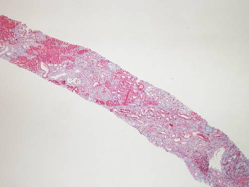
(THIS PICTURE BELOW IN LOW POWER SHOWS ONE OF THE CLASSICAL FINDINGS IN CSA TOXICITY)


A. Interstitial Fibrosis  
B. Tubular atrophy 
C. Endothelial cell swelling 
D. Glomerular thrombin deposits
E. Glomerular basement membrane thickening 
F. Double contours of the GB 
Incorrect
The correct answer is D.

Glomerular thrombin deposits

Explanation:

In a patient with suspected cyclosporine nephrotoxicity, renal biopsy reveals obliterative arteriolopathy (classically in the afferent renal arterioles), suggesting primary endothelial damage and subsequent endothelial cell swelling that may persist for months with elevated cyclosporine blood levels. This is also associated with a thickened glomerular basement membrane and a double-contour pattern. In fact, according to BANF, a thickened glomerular basement membrane and double-contour pattern most suggest chronic allograft nephropathy (CAN), also called transplant glomerulopathy. 

The other renal biopsy findings of cyclosporine nephrotoxicity include ischemic collapse or scarring of the glomeruli, vacuolization of the tubules, FSGS, and focal areas of tubular atrophy and interstitial fibrosis (producing a picture of "ZEB" A" or "STR "PED" fi" rosis). These changes occur with both low- and high-dose cyclosporine therapy, although they correlate earlier with higher doses. 

(THE ABOVE PICTURE IN THE UPPER HALF SHOWS TUBULAR ATROPHY APPEARING DARK AND REDDISH ALTERNATING WITH LIGHT BLUE AREAS OF INTERSTITIAL FIBROSIS GIVING A "STR "PED" R" "ZEB" A "APPEARANCE)

THE PICTURE BELOW SHOWS TUBULAR ATROPHY, VACUOLIZATION OF THE TUBULES AND ISCHEMIC CHANGES:


Mild arteriolar hyalinosis at six months appears to be associated with high doses of cyclosporine and was reversible. However, after more than a year, irreversible severe arteriolar hyalinosis and glomerulosclerosis were observed, despite decreased doses and trough levels of cyclosporine.

Glomerular thrombin deposits are typically seen in patients with Lupus, antiphospholipid syndromes and other vasculitides. It is typically not seen in cyclosporine nephrotoxicity.

Copyright © ABIM Exam World
Created On: 09/14/2017
Last Modified: 10/17/2026

End

Previous Next
Multiple
  • Test Id: 198396adfe94861c89
  • QId: 167347
  • 8 of 96
  • Notes
  • Calculator
  • Feedback

A 68-year-old African American male with a known history of diabetes mellitus,  hyperlipidemia, CVD, and GERD presents to the ER with chest pain and shortness of breath. The patient says the pain is crushing and radiates to his left arm. It woke him from sleep 30 minutes ago. He took his antacids and 4 tablets of nitroglycerin, and the pain got better. His current medications include low-dose aspirin, metoprolol, glyburide, pioglitazone, lisinopril, simvastatin, and omeprazole. He recently suffered an upper respiratory infection for which he was given Levofloxacin. Vitals: BP 160/95 mm Hg, T 98.1, HR 110. He has no leg edema and no jugular venous distention. EKG shows LVH by voltage criteria with an isoelectric ST segment. Serial troponin levels are normal. A stress test showed evidence of stress-induced ischemia in the anterior leads. He is scheduled for cardiac catheterization the next day. Laboratory results are as follows:

Serum Chemistry:

Na   143 mEq/L

K   3.5 mEq/L

Cl   101 mEq/L

HCO3  22 mEq/L

BUN   35 mg/dL

Cr   3.0 mg/dL

Glucose  110 mg/dL 

What would you do to PROTECT the kidneys before doing the cardiac catheterization?

A. Dopamine IV infusion before and after the  procedure.
B. Nifedipine.
C. Isotonic sodium bicarbonate infusion before and after the procedure.
D. N Acetyl Cysteine before and after the procedure
E. Start mannitol IV before the procedure
F. Left ventriculogram quickly with coronary angiogram
Correct
The correct answer is C.

Isotonic sodium bicarbonate infusion before and after the procedure. N-acetylcysteine is also used, but its efficacy in studies is unproven.

Explanation:

Contrast nephropathy usually presents as a rise in creatinine 24-48 hrs after IV radiocontrast exposure.

Other features of CONTRAST NEPHROPATHY are:
  • Low fractional excretion of sodium (FeNa) below 1%.
  • Iodinated contrast agents can falsely elevate protein so that proteinuria may be overestimated.d
  • It is reversible most of the time.
  • UA may show muddy brown granular casts, tubular epithelial cell casts and numerous tubular epithelial casts.

Contrast-associated nephropathy has to be differentiated from atheroembolic disease, which presents with deterioration in kidney function days to weeks after the procedure,  embolic lesions (such as digital ischemia of the toes) or livedo reticularis, transient eosinophilia and hypocomplementemia,  protracted course with frequently little or no recovery of renal function, and kidney biopsy showing the typical biconvex, needle-shape" cleft" ("ghosts") within the occluded vessel from the space occupied by the cholesterol crystal that dissolves during the processing of the tissue'giving a "ish mouth' appearance.

(Choice A) No evidence supports dopamine infusion as beneficial. In fact, it may be harmful.

(Choice B) No evidence supports that nifedipine is beneficial.

(Choice C) Studies suggest that infusion of isotonic saline or half-normal saline may be beneficial. Since alkalization may protect against free radical injury, sodium bicarbonate may be superior to isotonic saline. In patients who tolerate extra volume (i.e., those who are not volume overloaded or those who are not in heart failure), it would be reasonable to infuse isotonic sodium bicarbonate or saline.

(Choice DSomee studies suggest thatoral N-acetylcysteinee before and after theprocedure reducese the risk of contrastnephropathyl. Other studies suggest it is not beneficial. N-acetylcysteine is believed to work through its vasodilator and antioxidant effects. Because N-acetylcysteine is safe and inexpensive, many physicians would administer it prophylactically as well, even though results are conflicting. 

(Choice E) There are studies to suggest that prophylactic mannitol or furosemide is harmful.
    
(Choice F) Left ventriculogram should be avoided in patients at high risk of developing contrast nephropathy to minimize the quantity of IV contrast needed.

RISK FACTORS for contrast nephropathy are :
  • Chronic kidney disease
  • Diabetes mellitus
  • Congestive heart failure
  • Myeloma kidney
  • Hypotension

Educational Objective:

1. Identify and diagnose contrast nephropathy

2. Prevention of contrast nephropathy

Copyright © ABIM Exam World
Created On: 09/14/2017
Last Modified: 10/17/2026

End

Previous Next
Multiple
  • Test Id: 198396adfe94861c89
  • QId: 16521
  • 9 of 96
  • Notes
  • Calculator
  • Feedback

As per the JNC VIII committee recommendation, for individuals who are part of the African American population, including those with diabetes, the initial treatment should include a thiazide-type diuretic or calcium channel blocker (CCB).  

Which of the following thiazide-type diuretics is preferred as the initial antihypertensive therapy?


A. Hydrochlorthiazide 
B. Chlorthalidone 
C. Indapamide 
D. Metalazone 
E. Polythiazide 
Correct
The correct answer is B 
Chlothalidone

Explanation:

• Chlorthalidone at the same dose is approximately 1.5 to 2 times as potent as hydrochlorothiazide.

• Chlorthalidone has a longer duration of action than hydrochlorothiazide.

• There are no randomized clinical trials that directly compare outcomes in hypertensive patients treated with hydrochlorothiazide versus chlorthalidone. A multiple treatment (network) meta-analysis of nine trials including 50,946 patients was conducted in which hydrochlorothiazide and chlorthalidone were indirectly compared by evaluating their efficacy against common comparative drugs (ACE inhibitors were compared with hydrochlorothiazide in the ANBP2 trial and with chlorthalidone in the ALLHAT trial). The major findings of this meta-analysis showed that chlorthalidone significantly reduced the risk of cardiovascular events compared to hydrochlorothiazide (relative risk 0.79, 95% CI 0.72 to 0.88) and heart failure (relative risk 0.77, 95% CI 0.61 to 0.98). The authors calculated that 27 patients would need to be treated with chlorthalidone instead of hydrochlorothiazide over five years to prevent one cardiovascular event. Chlorthalidone remained superior to hydrochlorothiazide even after the meta-analysis was controlled for achieved office systolic blood pressure (relative risk for cardiovascular events 0.82, 95% CI 0.70 to 0.97). This finding may reflect chlorthalidone's longer duration of action and lower nocturnal blood pressure.

• Multiple Risk Factor Intervention Trial (MRFIT) - Men Hypertensive 2392 were treated with chlorthalidone, and 4049 were treated with hydrochlorothiazide. During six years of follow-up, cardiovascular events (defined as myocardial infarction, stroke, coronary artery bypass surgery, heart failure, left ventricular hypertrophy, peripheral artery disease, or angina) were significantly less common with chlorthalidone compared with hydrochlorothiazide (hazard ratio 0.79, 95% CI 0.68 to 0.92). Throughout the study, systolic blood pressure and LDL cholesterol levels were also lower with chlorthalidone than with hydrochlorothiazide. 

• Indapamide is a thiazide-like diuretic and has a half-life of 14-16 hours. This drug has been used in the HYVET trial alone or in combination with perindopril in the treatment of hypertension in patients more than 80 years old. The study showed a 30% reduction in stroke, a 39% reduction in the rate of death, 21% reduction in death from any cause, 23%reduction in CV death and 64%reduction in the rate of heart failure. The trial showed that careful BP lowering in the very elderly is beneficial. Indapamide is not preferred over chlorthalidone.

• Metolazone - There are many studies with metolazone available. An important additional property is its effectiveness as a diuretic at lower GFR values. The duration of action is about 24 hours.
It is not preferred over chlorthalidone.

Copyright © ABIM Exam World
Created On: 09/20/2017
Last Modified: 10/16/2026

End

Previous Next
Multiple
  • Test Id: 198396adfe94861c89
  • QId: 16524
  • 10 of 96
  • Notes
  • Calculator
  • Feedback

Which of the following antihypertensive medications resulted in better blood pressure control and cardiovascular outcomes when combined with an ACE inhibitor?


A. Beta blocker 
B. Calcium channel blocker
C. Angiotensin Receptor Blocker 
D. Diuretics 
Correct
The correct answer is B
Calcium channel blocker 

Explanation:

The ACCOMPLISH trial clearly showed that an ACE inhibitor combined with a Calcium channel blocker (amlodipine) is better than an ACE inhibitor combined with a diuretic (Hydrochlorothiazide). This study did not involve beta blockers or angiotensin receptor blockers. 

The ACCOMPLISH trial was the first major clinical trial addressing the issue of combination therapy in 11,000 patients who were at high cardiovascular risk (with prior CAD, diabetes, and/or CKD). The patients were randomly assigned to combination therapy with Benazepril (20 mg/day) combined with either amlodipine (5 mg/day) or Hydrochlorothiazide (12.5 mg/day).

The mean blood pressure was significantly lower in the Benazepril + Amlodipine group, and the primary endpoint was also significant in this group. The treatment combination of benazepril-amlodipine also showed a similar benefit on the secondary endpoint of cardiovascular death compared with the group treated with amlodipine and Hydrochlorothiazide. 

Copyright © ABIM Exam World
Created On: 09/20/2017
Last Modified: 10/16/2026

End

Previous Next
Multiple
  • Test Id: 198396adfe94861c89
  • QId: 165212
  • 11 of 96
  • Notes
  • Calculator
  • Feedback

The ACCOMPLISH trial is the first major trial addressing the issue of combination therapy in 11,506 patients who were at high cardiovascular risk. The goal blood pressure was <130/80 mm Hg in patients with diabetes or impaired renal function, and <140/90 mm Hg in patients with prior cardiovascular disease.

Which of the following combinations of blood pressure medications was the best in reducing cardiovascular events and slowing the progression of nephropathy in patients with hypertension who were at high risk for such events?

A. ACEI + Diuretics
B. ACEI + CCB 
C. ACEI + Beta-blocker 
D. CCB + Beta-blocker 
Correct
The correct answer is B
ACEI + CCB

Explanation:

• The Avoiding Cardiovascular Events through Combination Therapy in Patients Living with Systolic Hypertension (ACCOMPLISH) trial showed that initial antihypertensive therapy with benazepril plus amlodipine was superior to benazepril plus hydrochlorothiazide in reducing cardiovascular morbidity and mortality.

• ACCOMPLISH ACCOMPLISH was a double-blind, randomized trial undertaken in five countries (USA, Sweden, Norway, Denmark, and Finland). 11 506 patients with hypertension who were at high risk for cardiovascular events were randomly assigned via a central, telephone-based interactive voice response system in a 1:1 ratio to receive benazepril (20 mg) plus amlodipine (5 mg; n=5744) or benazepril (20 mg) plus hydrochlorothiazide (12.5 mg; n=5762), orally once daily. Drug doses were force-titrated for patients to attain recommended blood pressure goals.

• The primary endpoint was the composite of death from cardiovascular causes, nonfatal myocardial infarction, nonfatal stroke, hospitalization for angina, resuscitation after sudden cardiac arrest, and coronary revascularization.

• The benazepril-amlodipine combination was superior to the benazepril-hydrochlorothiazide combination in reducing cardiovascular events in patients with hypertension who were at high risk for such events.

Copyright © ABIM Exam World
Created On: 09/20/2017
Last Modified: 10/16/2026

End

Previous Next
Multiple
  • Test Id: 198396adfe94861c89
  • QId: 165215
  • 12 of 96
  • Notes
  • Calculator
  • Feedback

An 18-year-old male is brought to the renal clinic for evaluation of hypertension. There is no history of edema of the feet, puffiness of the face, hematuria, oliguria or recurrent urinary tract infections. He is an active football player, but lately he complains of weakness and muscle cramps. His blood pressure for the last 3-4 years has always been borderline high and during this visit was as noted below.

Physical examination shows: Pulse 100/min; all peripheral pulses are well felt. BP 150/100 mm Hg, Temp. 37.4 degrees C. He is pale. His growth is stunted. His cognitive function is normal, and no focal neurological deficits are noted. Other systems are unremarkable. Laboratory examination is as follows:

Hemoglobin   13.5 g/dL

Hematocrit   42%

Platelet Count   150,000 mm3

White Blood Cells  8,000 mm3


Urinalysis: 

Protein     100 mg/24 h

Glucose       None

Red Blood Cells      None

White Blood Cells     None

Leukocyte Esterase   Negative

Nitrites     Negative


Chemistry: 

BUN   13.5 mg/dL

Creatinine  0.9 mg/dL

Sodium   140 mEq/L

Potassium  5.8 mEq/L

Bicarbonate  18.5 mEq/L

Chloride   112 mEq/l

Calcium   9.2 mEq/L

Phosphorus  3.2 mg/dL

Aldosterone  5 ng/mL

PRA    <1.0 ng/dL/hr


Glucose   100 mg/dL

HbA1C    5.30%

S. Osmolality   282 mOsmol

Uric Acid   5.3 mg/dL

ESR    9.8 mm/h


ABG: 

pH   7.25

PCO2  32 mm Hg

HCO3 16 mEq/L

PO2   90 mm Hg

USG: Normal-sized kidneys, no hydronephrosis 

Considering the history and the laboratory findings, which of the following is most likely the cause of hypertension in this patient?


A. Liddle's Syndrome 
B. Gordon's Syndrome 
C. Congenital adrenal hyperplasia 
D. Glucocorticoid-remediable aldosteronism 
E. Mineralocorticoid receptor activating mutation 
Correct
The correct answer is B
Gordon’s syndrome

Explanation:

Gordon’s syndrome - A young male with hypertension, hyperkalemia, hyperchloremic metabolic acidosis, normal renal function, low renin, and low aldosterone is highly characteristic of Gordon’s syndrome. This syndrome is also known as pseudohypoaldosteronism type II. Recently, it has been reported that Gordon’s syndrome is linked to chromosomes 1, 12, and 17. Hyperkalemia and hypertension in Gordon’s syndrome are caused by abnormalities in WNK kinases, proteins that are localized to the distal nephron and affect the thiazide-sensitive Na-Cl Cotransporter. Mutations in WNK4 or WNK1 increase chloride reabsorption in the distal nephron, reducing lumen electronegativity and lowering the driving force for potassium secretion. These mutations also decrease expression of the potassium channels through which potassium enters the collecting duct. Thus, mutant WNK4 increases sodium chloride reabsorption in the distal tubule and decreases potassium secretion in the collecting duct, resulting in sodium retention, decreased potassium excretion, hypertension, hyperkalemia, and metabolic acidosis. Volume expansion suppresses renin and aldosterone. 

(Choice A) Liddle’s syndrome presents as hypertension in childhood. It is usually resistant to standard antihypertensive drugs. This is due to a gain-of-function mutation on chromosome 16 affecting the beta or gamma subunit of the ENaC channel in the collecting duct of the nephron. This results in sodium retention and volume expansion, causing hypertension and hypokalemia. Volume expansion suppresses renin. These patients will have hypertension, hypokalemia, metabolic alkalosis, low renin and low aldosterone. 

(Choice C) Congenital adrenal hyperplasia presents in childhood with hypertension and virilization in girls and precocious puberty in boys. The other features include primary amenorrhea, delayed sexual maturation, ambiguous genitalia, with clitoral enlargement in girls. Deficiencies in 11-beta hydroxylase (CYP11B1) and 17-alpha hydroxylase (CYP17) lead to excess deoxycorticosterone, which causes hypertension through mineralocorticoid action. Biochemically, it is characterized by metabolic alkalosis, low renin and low aldosterone.

(Choice D) Glucocorticoid-remediable aldosteronism (GRA) is also known as familial hyperaldosteronism type I. In this disorder, a chimeric gene forms from portions of the 11-beta-hydroxylase gene (CYP11B2) and the aldosterone synthase gene (CYP11B2). Patients with GRA have ectopic aldosterone synthesis occurring in the zona fasciculata under the direct influence of ACTH rather than renin. GRA presents with hypertension, hypokalemia, metabolic alkalosis, low renin and high aldosterone. GRA resembles a primary hyperaldosteronism state with excessive urinary potassium loss. Look for a clue of family history of CVA or brain haemorrhage in the stem of the question.

(Choice E) A mineralocorticoid receptor-activating mutation is a very rare autosomal dominant disorder. The mutation lies in the mineralocorticoid receptor, causing it to be activated by steroids lacking 21–hydroxyl groups, such as progesterone and spironolactone. Both normally antagonize the receptor but have opposite effects on this mutant receptor. It usually presents as early-onset hypertension in females. It may get accelerated during pregnancy due to elevated levels of progesterone.Biochemically, it is characterized by hypokalemia, metabolic alkalosis, o renin and low aldosterone.

Educational objective:

IF YOU KNOW THESE 5 POINTS FOR THESE 55 SYNDROMES THEN YOU CAN ANSWER ALL NEPHROLOGY BOARD QUESTIONS ASKED ON THESE SYNDROMES:

Blood pressure - Normotension or hypertension
Potassium        - Hyperkalemia or hypokalemia
Acid-Base         - Acidosis or alkalosis
Renin                - Low or High
Aldosterone      - Low or High

Copyright © ABIM Exam World
Created On: 09/20/2017
Last Modified: 10/16/2026

End

Previous Next
Multiple
  • Test Id: 198396adfe94861c89
  • QId: 165230
  • 13 of 96
  • Notes
  • Calculator
  • Feedback

A 56-year-old male was brought to the emergency room with drowsiness and lethargy. He has had these symptoms for the past 2 days. He complains of a recent history of anorexia, nausea, and vomiting. He has diabetes mellitus and has been on glimepiride 1 mg daily for the last 4 years. One week ago, he had decreased vision with redness in his right eye. He was treated by his ophthalmologist with drops,s which seem to have resolved the problem. He currently takes cholecalciferol weekly for osteoporosis. On physical examination, his pulse is 80/min, blood pressure is 140/90 mm Hg, respiratory rate is 20/min, and temperature is 97.7°F. The patient appears drowsy but shows no focal neurological deficits. Review of systems is otherwise unremarkable. Urinalysis is positive for glucose and negative for proteinuria, WBCs, and RBC casts. A 24-hour urinary protein collection shows proteinuria of 3.5 g/day. Further labs reveal:

Hemoglobin  8 gm%

Hct    24%

MCV   85

WBC   7800/ml

PMN   80%

Lymphocytes  20%

ESR   80 mm/hr

Na    145 mEq/L

BUN   80 mg/dL

Cr    1.8 mg/dL

CL    115 mEq/L

HCO3   25 mEq/L

Uric acid   5.8 mg/dL

Ca    14 mg/dl

PO4    2.8 mg/dL

Total Protein  7.8 gm/dL

Albumin   3.5 mg/dL.

Vitamin D  40 ng/ml

PTH   10 pg/ml

Which of the following is most likely the cause of his hypercalcemia?


A.  Vitamin D toxicity
B. Primary hyperparathyroidism
C.  Multiple myeloma.
D. Sarcoidosis
Correct
The correct answer is C.

Multiple myeloma

Explanation:

Multiple myeloma results from the proliferation of plasma cells producing a monoclonal immunoglobulin.

The diagnosis of multiple myeloma is supported by the presence of many of the following factors:
  • Advanced Age
  • Backache and Bone pain
  • Anemia
  • High ESR
  • Urine protein negative by dipstick and positive by other tests is characteristic of light chain proteinuria.
  • High Total protein
  • Low anion gap
  • Hypercalcemia and hyperuricemia favor the diagnosis of multiple myeloma.

(Choice A) This patient has received Vitamin D treatment. Vitamin D toxicity can give rise to hypercalcemia. Serum vitamin D3 levels will be high. PTH will be low. Patient’s vitamin D level of 40ng/ml (normal range is 30-100ng/ml) does not favor Vitamin D toxicity.

(Choice B) Primary hyperparathyroidism is the commonest cause of hypercalcemia among outpatients. It is characterized by elevated PTH. The patient’s PTH level of 10 pg/ml rules out primary hyperparathyroidism.

(Choice D) Sarcoidosis is a multisystem granulomatous disorder of unknown cause. Hypercalcemia and hypercalciuria in sarcoidosis are associated with high vitamin D levels. This patient has low vitamin D levels, so it is unlikely to be the cause. There is no history or findings to suggest sarcoidosis.

Copyright © ABIM Exam World
Created On: 09/20/2017
Last Modified: 10/17/2026

End

Previous Next
Multiple
  • Test Id: 198396adfe94861c89
  • QId: 165233
  • 14 of 96
  • Notes
  • Calculator
  • Feedback

A 25-year-old female is referred by her primary care provider for evaluation of hypertension and hypokalemia. Her primary care provider has already started her on oral potassium; despite this, her potassium remains low. Despite amlodipine and lisinopril, her blood pressure is 154/96 mm Hg. There is no renal bruit. Systemic and fundus examinations are normal. Her mother was also diagnosed with hypertension at an early age. Her brother died of a cerebrovascular accident 2 years ago. Laboratory findings are as follows:

Na   140 

Potassium 2.8 

Chloride   100 

HCO3    26

BUN   15 

Creatinine  0.8 

Glucose    110

TSH and Cortisol are normal

ACTH    elevated

Renin   0.7 (Low)

Aldosterone  48 (elevated)


Urinalysis:

Sodium   240 mEq/D

Potassium  98 mEq/D

Urinary 18-OH Cortisol and 18-oxocortisol are elevated.

The most appropriate treatment for this patient is:

A. Steroids
B. Spironolactone 
C. Steroids + Spironolactone
D. Amiloride 
Correct
The correct answer is C.

Steroids + Spironolactone

Explanation:

This patient has (GRA), glucocorticoid-responsive aldosteronism. These are typically young patients presenting with hypertension and hypokalemia. There is a strong family history of early-onset hypertension along with a history of fatal CVA or hemorrhagic strokes. These patients typically have low Renin and high aldosterone, mimicking a state of primary hyperaldosteronism. As a result, they have high urinary potassium losses, as seen in this patient. (Urinary potassium of more than 40mEq/D is considered increased losses). Increased ACTH and 18-OH cortisol further support the diagnosis of GRA.

Steroids alone are used in the treatment of pregnancy-associated mineralocorticoid excess and congenital adrenal hyperplasia. Steroids alone in this patient will suppress ACTH, but this patient also needs an aldosterone antagonist for the hyperaldosteronism and to reduce urinary potassium losses. Hence, the right answer is steroids plus spironolactone.

Spironolactone alone is not sufficient, as steroids are needed to suppress the increased ACTH state. 

Amiloride blocks the ENaC channel in Liddle syndrome.

PLEASE NOTE THAT "G" AND "S" ARE FREQUENTLY ASKED QUESTIONS IN NEPHROLOGY BOARD EXAMS

PLEASE REVIEW GLUCOCORTICOID-REMEDIABLE ALDOSTERONISM WELL. 

Copyright © ABIM Exam World
Created On: 09/20/2017
Last Modified: 10/17/2026

End

Previous Next
Multiple
  • Test Id: 198396adfe94861c89
  • QId: 165242
  • 15 of 96
  • Notes
  • Calculator
  • Feedback

A 60-year-old with recently diagnosed colon cancer and diabetes presents with bilateral pedal edema; BP is 120/80 mm Hg. Urinalysis showed  4+ protein, no RBCs or WBCs, and 8-10 Hyaline casts. His BUN is 20, Creatinine is 1 mg/dL, and albumin is 2 grams/dL. A 24-hour urine collection showed 10 grams of protein. The patient undergoes a kidney biopsy. The EM is shown below :


What is the most likely diagnosis?

A.  Crescentic GN
B.  IgA nephropathy
C.  Minimal change disease
D.  Focal segmental glomeruloscerosis
E.  Membranous nephropathy
F. Diabetic nephropathy
Correct

The correct answer is E

Membranous Nephropathy.

Electron microscopy shows subepithelial electron-dense deposits, as classically seen in membranous nephropathy. If in the question there is a suggestion of colon, breast, or lung cancer, then the glomerulopathy is usually membranous. Therefore, histopathological findings support the diagnosis: subepithelial electron-dense deposits.

Explanation:

BOARD POINT - FAMILIARIZE YOURSELF WITH THESE ASSOCIATIONS :

1. Solid cancers (colon, breast, lung, renal) plus proteinuria = Membranous nephropathy

2. Hodgkin's lymphoma plus proteinuria = Minimal change disease

3. HIV plus proteinuria = Focal segment glomerulosclerosis FSGS

4. Pamidronate plus protenuria = FSGS (rare)

5. Myeloma, no albuminuria on dipstick, but proteinuria on protein/creatinine ratio in 24-hour urine: Light chain nephropathy

6. Myeloma th with nonspecific proteinuria (dipstick, urine protein/creatinine ratio in 24-hour urine): Light chain nephropathy or amyloidosis.


BOARD POINT - FAMILIARIZE YOURSELF WITH THESE HISTOPATHOLOGY ASSOCIATIONS FOR VARIOUS GLOMERULAR DISEASES


Copyright © ABIM Exam World
Created On: 09/20/2017
Last Modified: 10/17/2026

End

Previous Next
Multiple
  • Test Id: 198396adfe94861c89
  • QId: 165243
  • 16 of 96
  • Notes
  • Calculator
  • Feedback

You are the nephrologist on call. The  ER calls you for an 18-year-old female who is complaining of vomiting and diarrhea. Her serum sodium is 116 mEq/L and Serum potassium is 5.9 mEq/L. On physical examination, the patient is drowsy; pulse 126/min, BP 90/60 mm Hg, and RR 32/min. Her chest is clear. Her heart sounds are normal, with no murmur. The patient is drowsy but arousable, with no focal neurological deficit. Laboratory findings are as follows:

Hb    16 gm/dl 

WBC   12,800/cmm 

Polymorph  46% 

Lymphocytes  16% 

Eosinophils  4%

Monocytes  4%

Platelets   2,40,000/cmm.

CL    70 mEq/L

BUN  10 mg/dl

Creatinine  0.5 mg/dl

Na    116 mEq/L

K    5.8 mEq/L


Urinalysis:

pH    6.4

Protein  trace

Glucose  absent

Microscopic: occasional WBCs & RBCs

Urinary Na  90 mEq/L

Urinary K         20 mEq/L

ABG    

PH                   7.32

PCO2   36 

HCO3   20 mEq/L

PaO2   92

O2 saturation  98%

S. Cortisol  6.00 mg/dl

TSH   3.5 IU/m (Normal 0-5 IU/m ).

Both plasma Renin and Aldosterone are high. 

Which of the following conditions is most likely with these findings?


A. Type 4 RTA 
B. Pseudo-hypo-aldosteronism Type 1 
C. Gordon's syndrome
D.  Diarrhea
Incorrect
The correct answer is B.
 Pseudo-hypoaldosteronism type 1

Explanation:

This patient has hyponatremia with high urinary sodium, high serum potassium, low urinary potassium, and normal adrenal function. The serum aldosterone level is high, suggesting target-organ resistance to aldosterone. These findings are characteristic of pseudohypoaldosteronism type 1 (PHA type 1)

PHA type 1 is a rare hereditary disorder characterized by generalized resistance to aldosterone. It presents with 
1. Salt wasting 
2. Hypovolemia 
3. Normotension
4. Metabolic acidosis 
5. Hyperkalemia
6. High Renin and Aldosterone.  

NEPHROLOGY BOARD EXAM TAKERS REMEMBER THESE 6 POINTS FOR PHA TYPE 1

These are two different modes of inheritance:
1] Autosomal recessive.
2] Autosomal dominant.

Autosomal recessive affects the epithelial sodium channel and other target organs, such as the kidney, colon, and sweat gland. This causes downregulation of sodium channels and decreased sodium transport.

The autosomal dominant or sporadic form is due to heterozygous mutations in the NR3C2 gene, which codes for the mineralocorticoid receptor. This is a milder form than autosomal recessive disease, in which only the kidney is affected. The disease often improves with age.

Treatment consists of a high-salt diet. This prevents volume depletion and, by enhancing sodium delivery to the distal tubules, increases potassium excretion, thereby lowering serum potassium. High-dose fludrocortisone (1 to 2 mg/d), or carbenoxolone, is indicated if high-salt intake is ineffective or not tolerated.

(Choice A) Type IV RTA has low aldosterone and low renin levels; therefore, this is not the likely answer.

(Choice D) Diarrhea will have metabolic acidosis with hypokalemia, and Net urine charge will be negative. This patient has positive Net urine charge with hyperkalemia, which rules out diarrhea as a cause.

(Choice C)  = PHA TYPE 2 (Pseudohypoaldosteronism type 2) OR = Gordon’s syndrome is characterized by:
 1. Hypertension 
 2. Hyperkalaemia 
 3. Metabolic acidosis
 4. Low plasma Renin and Aldosterone. This is due to mutations in WNK kinases 1 and 4. These mutations increase chloride reabsorption and sodium retention, resulting in hypertension.

NEPHROLOGY BOARD EXAM TAKERS REMEMBER THESE 4 POINTS FOR PHA TYPE 2 OR GORDOGORDON'S ROME

IF YOU KNOW THESE 10 POINTS OF PSEUDOHYPOALDOSTERONISM TYPE 1 & 2 THA, THEN YOU CAN ANSWER ALL QUESTIONS ON PHA 1, 2 AND GORDON'S SYNDROME, WHICH ARE GOING TO BE ASKED FOR SURE ON THE BOARDS.

Copyright © ABIM Exam World
Created On: 09/20/2017
Last Modified: 10/17/2026

End

Previous Next
Multiple
  • Test Id: 198396adfe94861c89
  • QId: 165246
  • 17 of 96
  • Notes
  • Calculator
  • Feedback

A 32-year-old male is brought to the renal clinic with a history of hematuria, oedema of the feet, and puffiness of the face. He reports fever and sore throat a week ago. He also complains of breathlessness on exertion and oliguria. Physical examination shows: Pulse 100/min, BP 150/100 mm Hg, and Temp. 37.4 C. He is pale. He has facial puffiness and oedema of the feet. Systemic examination-unremarkable. Laboratory examination is as follows:

Hb   10.5 g/d

Hct   34%

Platelet 250,000 mm3

WBC  8,000 mm3

Differential count: P 80% L 12% E 6% M 2%

ESR  9.8 mm/h


Urinalysis:

Protein   3000 mg/24 h

Glucose   None

RBC   50-60/hpf Dysmorphic

WBC   occasional

Leukocyte Esterase Negative

Nitrites Negative

BUN   40 mg/dL

Creatinine  3.9 mg/dL

Sodium   140 mEq/L

Potassium  4.2 mEq/L

Bicarbonate  25.5 mEq/L

S. protein  5.5 g/dl

S. Albumin  2.5 g/dl

Calcium   9.2 mEq/L

Phosphorus  3.2 mg/dL

Glucose   100 mg/dL

Uric Acid   5.3 mg/dL

C 3    Low

C4     normal

HBsAg /HIV   Neg

ANA    Neg

Kidney Biopsy: Shows enlarged Glomeruli, lobular accentuation, mesangial hypercellularity, endo-capillary proliferation and double contour along the capillary wall. IF shows bright C3 in the mesangium and capillary wall with absent immunoglobulin staining. 

Electron Microscopy: Suggestive of dense deposits.

What is the BEST treatment option for this patient?


A. Plasma exchange + Rituximab 
B. Rituximab 
C. Eculuzimab
D. Cyclophosphamide + Steroids 
Correct
The correct answer is C.

Eculuzimab

Explanation:

This patient has a nephritic-nephrotic picture with low C3. The Kidney biopsy, along with the clinical presentation, suggests MPGN. Negative immunoglobulins, along with positive C3 staining, narrow it down to DDD (DENSE DEPOSIT DISEASE) or C3-GN (C3 glomerulonephritis)

DDD, or dense deposit disease, is best treated with Eculizumab. 
Rituximab is not useful in DDD or C3-GN. 
ECULUZUMABB:is usefull in:
1. DDD
2. Atypical HUS – used along with plasma exchange. If using eculizumab, give the meningococcal vaccine or give penicillin till the vaccine becomes effective.

However, (additional information not about this question)

RITUXIMAB: is useful in:
1. ANCA vasculitis (can be used in induction or relapse – RAVE TRIAL)
2. Wegener's
3. HCV cryoglobulinemia
TTP

***** TREATMENT OPTIONS IN GLOMERULONEPHRITIS WITH RITUXIMAB AND ECULIZUMAB ARE FREQUENTLY TESTED CONCEPTS IN NEPHROLOGY BOARD EXAMS. PLEASE REVIEW THESE IN DETAIL.*****


Copyright © ABIM Exam World
Created On: 09/20/2017
Last Modified: 10/17/2026

End

Previous Next
Multiple
  • Test Id: 198396adfe94861c89
  • QId: 165247
  • 18 of 96
  • Notes
  • Calculator
  • Feedback

A 36-year-old female was diagnosed with membranous nephropathy secondary to SLE. Her 24-hour protein excretion was 7.5 gms/day. Her serum creatinine was 0.9mg/dl. She was started on 500 mg of cyclophosphamide IV every 15 days (Euro-Lupus) and prednisolone 1 mg/kg orally per day. After 3 months of therapy, she presented with decreased urine output, puffiness of the face, and oedema of the feet. On physical examination, her temperature was 37°C, blood pressure was 160/100 mm Hg, pulse was 90/min, and respiratory rate was 20/min. She is anemic, with facial puffiness and foot oedema. On systemic examination, air entry was decreased at the bases of both lung fields, and heart sounds were distant and feeble. Chest X-Ray reveals bilateral pleural effusions. Echocardiogram reveals mild to moderate pericardial effusion. Laboratory examination is as follows: 

Hemoglobin   10.0 g/dL

Hematocrit   34%

Platelet Count   150,000 mm3

WBC    8,000 mm3

Differential count: T P  80% L 12% E 6% M 2%

ESR    50.8 mm/h


Urinalysis: 

Protein  1450 mg/24 h

Glucose  None

RBCs  70-80/HPF dysmorphic

WBCs  5-8/HPF

Leukocyte Esterase Negative

Nitrites  Negative

 BUN   35 mg/dL

Creatinine  3.9 mg/dL

Sodium   140 mEq/L

Potassium  5.2 mEq/L

Bicarbonate  15.5 mEq/L

Calcium   9.2 mEq/L

Phosphorus  5.6 mg/dL

Glucose   100 mg/dL

Uric Acid   5.3 mg/dL

C3 & C4 decreased 

ANA   positive

dsDNA   positive

Repeat biopsy shows:


Which of the following is the most appropriate therapy for her current condition?


A. Mycophenolate and steroid
B. Mycophenolate, tacrolimus and steroids 
C. Rituximab 
D. I.V Immunoglobulins 
E. Plasma exchange  
Correct
The correct answer is B.

Mycophenolate, tacrolimus and steroids 

Explanation: 

This patient has membranous lupus being treated with the Euro-Lupus regimen. Urine activity, plus positive ANA and dsDNA and low C3 and C4, suggests disease activity. The biopsy shows active lupus nephritis combined with features of class IV & V diffuse proliferative GN. Patients with both DPGN and membranous lesions are more likely to be resistant to standard induction regimens with cyclophosphamide or MMF with steroids. In these patients, combined treatment with MMF and tacrolimus is recommended. This was suggested by Bao H. et al. in a small, short-term prospective trial in which 40 patients with diffuse proliferative plus membranous lupus nephritis were randomly assigned to induction therapy with MMF (0.75g to 1g/d) and tacrolimus (3-4 mg/d) or intravenous cyclophosphamide alone. All patients received steroids. At nine months, there was a significantly higher rate of complete remission in patients treated with MMF and tacrolimus as compared to cyclophosphamide (65% versus 15%).

(Choice A) Mycophenolate and steroids can be used in patients who have received cyclophosphamide and are resistant to it.

(Choice C and D) Rituximab and I.V. Immunoglobulins can be used in patients with lupus nephritis who have failed to respond to cyclophosphamide, MMF and steroids.

(Choice E) Plasma exchange is recommended in patients with SLE and(TT). 

KDIGO Clinical Practice Guidelines for Glomerulonephritis recommend the following treatment for resistant lupus nephritis:

Treatment of resistant disease 12.9.1: In patients with worsening S. Cr and/or proteinuria after completing one of the initial treatment regimens, consider performing a repeat kidney biopsy to distinguish active LN from scarring. (Not Graded) 12.9.2: Treat patients with worsening S. Cr and/or proteinuria who continue to have active LN on biopsy with one of the alternative initial treatment regimens. If the patient has received cyclophosphamide, use MM; if the f patient has received MMF, use cyclophosphamide (Not Graded) 12.9.3 

KDIGO suggests that non-responders who have failed more than one recommended initial regimen (cyclophosphamide, MM, or CNIs) may be considered for treatment with rituximab, IV Immunoglobulins, or CNIs. (2D).


Copyright © ABIM Exam World
Created On: 09/20/2017
Last Modified: 10/17/2026

End

Previous Next
Multiple
  • Test Id: 198396adfe94861c89
  • QId: 165256
  • 19 of 96
  • Notes
  • Calculator
  • Feedback

A 30-year-old man comes to your office for a painful rash on the neck. He has fever and malaise. He has a history of HIV. He is currently taking Tenofovir, emtricitabine, and indinavir. The rash is suggestive of Herpes Zoster rash :

Physical examination reveals no oral cavity lesions. His cu rent CD4 count is 250/mm3. Hischemistry iss normal. He is started on an intravenous medication for his rash. Two days later, his chemistry is as follows:

Na    135 mEq/L

K     4.5 mEq/L                  

CL    100 mEq/L

HCO3   24 mEq/L                  

BUN  21 mg/dL                       

Cr   2.0 mEq/L

Glucose  95 mg/dL

Calcium   9.4 mg/dl

Urinalysis shows needle-shaped crystals in the sediment.

Which of the following is most likely the cause of his renal problem?


A.  Indinavir
B.  Tumor lysis syndrome
C.  Antifreeze ingestion
D.  Acyclovir
E.  IV TMP/SMX
Correct
The correct answer is D.

Acyclovir

Explanation:

This patient has a characteristic herpes zoster lesion on his neck. The vesicles are 2-3 mm in size with an erythematous base. They are in different stages of development. Herpes zoster is commonly seen in the elderly and immunocompromised. This patient is 30 years old and immunocompromised (HIV positive with AIDS). The treatment of choice for herpes zoster in an immunocompromised patient is IV acyclovir. High-dose acyclovir is one of the causes of crystal-induced nephropathy. The crystals are needle-shaped.

Indinavir (Choice A), a protease inhibitor, is a common cause of nephrolithiasis in HIV patients. Patients would often present with flank pain. Urinalysis would show hematuria and needle-shaped crystals. This patient, however, had normal serum chemistry on presentation. Following two days of IV medication (acyclovir), his serum chemistry showed elevated creatinine, suggesting acyclovir as the most likely cause.

Tumor lysis syndrome (Choice B) occurs in the setting of chemotherapy for lymphoma.  It leads to the formation of needle-shaped uric acid crystals. This patient has no such presentation.

Patients with Antifreeze ingestion (Choice C) present with metabolic acidosis with an elevated anion gap. Initial urinalysis shows envelope-shaped oxalate crystals.

Trimethoprim-sulfamethoxazole (TMP/SMX) (Choice E) has sulfonamide in it. Sulfonamide also leads to crystal-induced nephropathy. The crystals are often dumbbell-shaped.

Educational Objective:

Drugs causing Crystal-induced nephropathy:

ACYCLOVIR --- Needle-shaped crystals

INDINAVIR --- Needle-shaped crystals

SULPHONAMIDE --- Dumbbell-shaped crystals

CALCIUM OXALATE (Antifreeze- Antifreeze-shapedd crystals

CALCIUM PO4---- Coffin-shaped crystals

URIC ACID --- Hexagonal crystals

CYSTEINE --- Hexagonal crystals


Copyright © ABIM Exam World
Created On: 09/20/2017
Last Modified: 10/17/2026

End

Previous Next
Multiple
  • Test Id: 198396adfe94861c89
  • QId: 165258
  • 20 of 96
  • Notes
  • Calculator
  • Feedback

 In patients with CKD and ESRD, Vitamin D appears to protect against cardiovascular events by controlling secondary hyperparathyroidism. The OPERA trial evaluated the effect of the Vitamin D analog paricalcitol in CKD stages 3-5 with left ventricular hypertrophy. The study observed the effect on left ventricular mass and function.

Which of the following statements is true about the effect of paricalcitol in CKD and ESRD patients?

A. Paricalcitol treatment reduced LV mass
B. Paricalcitol treatment preserved LV ejection fraction
C. Paricalcitol treatment improved pulse wave velocity and arterial stiffness
D. Paricalcitol treatment improved blood pressure control
E. Paricalcitol treatment significantly reduced PTH and alkaline phosphatase 
Correct
The correct answer is E

Paricalcitol treatment significantly reduced PTH and alkaline phosphatase.

Explanation:

Vitamin D Rx DOES NOT reduce LV mass OR LV ejection fraction OR arterial stiffness.s
Vitamin D seems to protect against cardiovascular disease, but the reported effects of vitamin D on patient outcomes in CKD are controversial. The 

OPERA trial was a prospective, double-blind, randomized, placebo-controlled trial to determine whether oral activated vitamin D reduces left ventricular (LV) mass in patients with stages 3- 5 CKD and LV hypertrophy.
Subjects with echocardiographic criteria of LV hypertrophy were randomly assigned to receive either oral paricalcitol (1 ?g) one time daily (n=30) or matching placebo (n=30) for 52 weeks.

The primary endpoint was change in LV mass index over 52 weeks, which was measured by cardiac magnetic resonance imaging. Secondary end points included changes in LV volume, echocardiographic measures of systolic and diastolic function, biochemical parameters of mineral bone disease, and measures of renal function.
Change in LV mass index did not differ significantly between groups (median [interquartile range], -2.59 [-6.13 to 0.32] g/m(2) with paricalcitol versus -4.85 [-9.89 to 1.10] g/m(2) with placebo).
Changes in LV volume, ejection fraction, and tissue Doppler-derived measures of early diastolic and systolic mitral annular velocities, and ratio of early mitral inflow velocity to early diastolic mitral annular velocity did not differ between the groups.

Paricalcitol treatment significantly reduced intact parathyroid hormone (P<0.001) and alkaline phosphatase (P=0.001) levels as well as the number of cardiovascular-related hospitalizations compared with placebo.

52 weeks of treatment with oral paricalcitol (1 ?g one time daily) significantly improved secondary hyperparathyroidism but did not alter measures of LV structure and function in patients with severe CKD and ESRD.

OPERA and PRIMO are two recent randomized clinical trials in patients with CKD that evaluated paricalcitol for slowing the progression of left ventricular mass. Both of these trials were found to have negative results.

Copyright © ABIM Exam World
Created On: 09/20/2017
Last Modified: 10/17/2026

End

Previous Next
Multiple
  • Test Id: 198396adfe94861c89
  • QId: 165260
  • 21 of 96
  • Notes
  • Calculator
  • Feedback

You are rounding on your patients in the dialysis unit and see a 65-year-old gentleman with ESRD due to chronic interstitial disease. He also has a history of diet-controlled diabetes mellitus and hypertension. His other past medical history is significant for dyslipidemia, coronary artery disease, hypothyroidism, gout and depression. He has been hospitalized in the recent past for swelling and pain of his right great toe. He was seen by the foot doctor, a scan was done, and eventually the great toe had to be amputated. H has been on hemodialysis 3 times a week. His weekly Kt/V is 1.9. You are conducting the monthly blood work review for this patient. You note that his hemoglobin has been persistently low for the past few monthly blood draws. He is currently on 100 mcg of Darbepoetin weekly on dialysis. On enquiry, there is no history of blood loss in the form of hematemesis, melena, hematochezia or hemoptysis. His active medications include losartan, Atorvastatin, Calcitriol, multivitamin supplements, paroxetine, allopurinol and aspirin.

His pertinent blood work is as follows:

Test

Result

WBC

4500 cells /cumm

Hemoglobin

8.2 g/dL

Platelet count

450 thousand /cumm

Reticulocyte count

Normal

Iron

55 (range 50-150)

Total iron-binding capacity

250 g/dl (range 250-310)

Transferrin saturation

20%

Ferritin

1400 ng/ml (range 20-235)

Haptoglobin

400 mg/dl (range 83-267)

Lactate dehydrogenase

240 U/L (range 80-225)

Total bilirubin

1.0 mg/dl

Folate

7 ng/ml (range 1.8-9.0)

B12

500 pg/ml (range 200-800)


Peripheral blood smear.

Normal RBC morphology, few burr cells.


Which of the following is true about this patient’s anemia?


Omitted

Correct Answer: Option C: This patient has chronic inflammation, which contributes to his anemia.


Explanation:

10-15% of patients receiving erythrocyte-stimulating agents (ESAs) develop resistance. There are multiple reasons why ESRD patients develop resistance.


ESA resistance occurs due to the following reasons:

  1. Iron deficiency.

  2. Chronic inflammation.

  3. Under-dialysis.

  4. Hemolysis.

  5. Folate and B12 deficiency.

  6. Chronic blood loss.

  7. Anti-EPO antibodies.

  8. Pure red cell aplasia.

  9. Failed chronic renal allograft.

  10. ACEI/ARB.

  11. Aluminum overload.

  12. Hyperparathyroidism.

  13. Hematological disorders or malignancy.


Option A: Incorrect option. ESRD is associated with erythropoietin deficiency—the patient has already been initiated on ESA. There is no need to measure EPO levels. There is no evidence that measuring EPO levels is part of anemia management in aKD.

Option B: Incorrect option. ESRD is an inflamed state. In an inflammatory milieu, hepcidin production increases. The hepatic iron-regulatory hormone Hepcidin and its receptor, the cellular iron exporter ferroportin, form a feedback-regulated mechanism that maintains iron-transferrin concentrations, erythropoiesis and other functions, ensuring sufficient iron availability and preventing toxicity. In chronic kidney disease, inflammation and impaired renal clearance increase plasma hepcidin, inhibiting duodenal iron absorption and sequestering iron in macrophages. Hepcidin can cause systemic iron deficiency, reduced iron availability for erythropoiesis, and resistance to endogenous and exogenous erythropoietin.

Choice C: Correct option. Refer to explanation for option B.  He had swelling in the great toe; a foot doctor saw him, a bone scan was done, and subsequently the amputation: all suggestive of an infective etiology, probably osteomyelitis. There is a temporal relationship between the patient’s symptoms and underlying chronic inflammatory state.

The high ferritin also suggests an inflammatory state.

Choice D: Incorrect option. Pure red cell aplasia, a form of severe ESA hyporesponsiveness mediated by anti-erythropoietin antibodies, was first reported with certain formulations of Epoetin alfa but has now been reported with all commercially available forms of ESA. This syndrome presents with rapid-onset severe anemia (hemoglobin <7 g/dL), severe reticulocytopenia (reticulocyte count <10,000/? L), and marked elevations in serum ferritin (>1000 ng/mL) and transferrin saturation (>70%) due to low iron utilization. Pure red cell aplasia is unlikely given the absence of characteristic laboratory findings. Moreover, the patient did ot receive Epoetin alfa.


Choice E: Incorrect option. Under-dialysis leads to anemia due to the same mechanism mentioned earlier in option B. Under-dialysis worsens the uremic milieu, which in turn leads to an inflammatory state. This leads to anemia. The patient in this clinic vignette has been dialysed appropriately. His weekly Kt/V is 1.9, which is above the target goal of 1.7

Copyright © ABIM Exam World
Created On: 10/22/2018
Last Modified: 10/16/2026

End

Previous Next
Multiple
  • Test Id: 198396adfe94861c89
  • QId: 165261
  • 22 of 96
  • Notes
  • Calculator
  • Feedback

50-year-old female patient whose group B is being evaluated for kidney transplant surgery. She had ESRD secondary to analgesic nephropathy and has been on hemodialysis for the last 5 years. She has had multiple sensitization events in the form of 3 pregnancies and several blood transfusions. Her current calculated PRA against class I antigen is 97% and against class II antigen is 99%. She has been enrolled in the national highly sensitized recipient program.

Her husband, who is blood group matched, came forward as a potential kidney donor, but she had a positive Flow B and T-cell crossmatch against him. Single antigen bead assay demonstrated donor-specific antibodies against class II across DQB*15 and DPB*14. This transplant did not materialize as the patient declined the desensitization protocol. Now her younger brother comes forward as a potential donor. He is blood group A, and the flow B and T-cell crossmatch is negative, with no demonstrable donor-specific antibodies against this donor. The patient wants to know more about ABO-incompatible transplant.


Which of the following statements about the ABO incompatible transplant is correct?


A. Three-year graft survival is inferior to blood group compatible transplants.
B. The infectious and bleeding complications post ABOI kidney transplant are the same as blood matched kidney transplant.
C. All patients undergoing ABOI transplant need to undergo desensitization using IVIg, Plasma exchange, Rituximab irrespective of their donor/recipient pair Anti ABO titers for optimal outcomes.
D. C4d staining on protocol biopsies is common feature and does not necessarily mean an antibody mediated rejection process in the absence of allograft dysfunction.
Incorrect

Correct answer: Option D: C4d staining on protocol biopsies is a common feature and does not necessarily mean an antibody-mediated rejection process.


Explanation:


Choice A: Three-year graft survival is inferior to blood group-compatible transplants is incorrect. A comprehensive database analysis of 1420 ABOI living donor (LD) kidney transplants performed in 101 centers from 2005 to 2012 compared graft and patient survival with a matched cohort of ABO-compatible transplant recipients. Three-year graft and patient survival were ultimately identical. 1


Choice B: The infectious and bleeding complications post ABOI kidney transplant are the same as in blood-matched kidney transplant is also incorrect. Using USRDS and Medicare data from 2000–2007, 119 ABOI (non-A2 donor) transplant recipients were identified. Compared with ABO-compatible recipients, the risks of infectious and hemorrhagic complications were significantly higher, with a 2.2-fold higher risk of pneumonia, a 3.5-fold higher risk of wound infections, a 56% higher risk of pyelonephritis, and a nearly 2- fold higher risk of hemorrhage 2


Choice C: All patients undergoing ABOI transplant need to undergo desensitization using IVIg, Plasma exchange, Rituximab irrespective of their donor/recipient pair. Anti-ABO titers for optimal outcomes is also an incorrect answer. Historically, ABOI transplantation has been successful when performed after desensitization with plasmapheresis, intravenous Ig (IVIG), rituximab, and/or splenectomy to achieve ABO IgG antibody titersof  1:4. A recent publication demonstrated that these intensified treatments might not be necessary in donor/recipient pairs who have low-to-moderate titer ABO incompatibility 3


Choice D: C4d staining on protocol biopsies is a common feature and does not necessarily mean an antibody-mediated rejection process; in the absence of allograft dysfunction, this is the correct answer. C4d staining is not an uncommon feature seen in the protocol biopsies done in ABOI kidney transplant recipients. In the absence of allograft dysfunction, C4d staining has no clinical relevance and is part of graft accommodation.

Copyright © ABIM Exam World
Created On: 10/30/2018
Last Modified: 10/16/2026

End

Previous Next
Multiple
  • Test Id: 198396adfe94861c89
  • QId: 165262
  • 23 of 96
  • Notes
  • Calculator
  • Feedback

A 28-year-old Caucasian gentleman comes to the clinic for a follow-up visit. He has been found to have type 1 diabetes mellitus since the age of 12. His cousin has the same disease. He reports no symptoms. He has been using an insulin pump with insulin aspart. He monitors blood sugar with a flash glucose monitor and uses carbohydrate counting for boluses. He reports infrequent hypoglycemic episodes, particularly 2 hours post-lunch, but manages by himself. He exercises at least60 minutes per day. His vitals show BP of 118/6  mHg. His BMI is 23.2. System examination is unremarkable.


His laboratory investigations are reported as follows.


Characteristic

Value

Hemoglobin

14.2 gm/L 

WBC count

6.8 X 10³/cubic mm

Platelet count

241 X 103/cubic mm

Segmented Neutrophils 

Lymphocytes

Monocytes

Band neutrophils 

Eosinophils

Basophils

60%

36%

2%

0%

2%

0%

Sr Sodium

139 mEq/L

Sr Potassium

4.4 mEq/L

Sr Creatinine

0.6 mg/dL

eGFR using CKD-EPI

136.8 ml/min/1.73m2

Sr Bicarbonate

24 mEq/L

Sr Chloride

101 mEq/L

Total Bilirubin

1.0 mg /dL

AST

16 U/L

ALT

18 U/L

Sr Albumin

4.0 g/dL

HBA1C

7.9%

Sr Calcium

10 mg/dL

Urine dipstick

pH- 5.4

Albumin-nil

 no blood

 no WBCs

24-hour urinary protein 

76 milligrams/day


What is the MOST LIKELY incorrect statement regarding the hyperfiltration stage of Diabetic Kidney Disease in this patient?

A. Renal hyperfiltration is usually diagnosed when the GFR is more than 120 ml/min,which corresponds to a renal function that exceeds two standard deviation above mean GFR.
B. Renal hyper filtration usually precedes microalbuminuria in type 1 diabetes mellitus.
C. Renal hyper filtration is considered as a risk factor for future progression to chronic kidney disease (CKD) and end stage renal disease (ESRD) in type 1 DM.
D. eGFR equations like MDRD equation can be used predict hyper filtration.
Incorrect

The Correct Answer is Option D: eGFR equations like the MDRD equation can be used to predict hyperfiltration.


Explanation:


Supraphysiological elevation in GFR is observed early in the natural history of type 1 and type 2 diabetes mellitus due to glomerular hyperfiltration. Pathogenesis of hyperfiltration in diabetes is complex, with a prominent role for hyperglycemia and distorted insulin levels, especially in early diabetes and pre-diabetes. Dilatation of the afferent (pre-capillary) glomerular arteriole plays an important role in the hyperfiltration response, by raising both the intra-glomerular pressure and renal blood flow.


Direct measurement of GFR is usually required to detect hyperfiltration because estimation equations, such as the Modification of Diet in Renal Disease (MDRD), usually underestimate the true GFR when it is normal or above normal. 


Option A: A definite GFR cutoff is lacking. However, renal hyperfiltration is typically defined as a GFR between 120 mL/min and 150 mL/min/1.73 m2, or greater than 2 standard deviations above the mean GFR in normal, healthy individuals.


Option B: Hyperfiltration in diabetes precedes the onset of albuminuria and/or decline in renal function, and predisposes to progressive nephron damage by increasing glomerular hydraulic pressure


Option C: Hyperfiltration alone does not seem to fully explain adverse renal outcomes, as the risk for ESRD in transplant donors is very low. However, in type 1 diabetes, rapid GFR decline is associated with renal hyperfiltration and impaired GFR and may predict progressive DKD before loss of renal function.

Copyright © ABIM Exam World
Created On: 10/30/2018
Last Modified: 10/16/2026

End

Previous Next
Multiple
  • Test Id: 198396adfe94861c89
  • QId: 165263
  • 24 of 96
  • Notes
  • Calculator
  • Feedback

A 19-year-old African American woman comes to the clinic for a follow-up visit. She has been found to have type 1 diabetes mellitus since the age of 12. She has been using an insulin pump for the last 5 years. She reports no hypoglycemic symptoms and monitors blood sugar with a flash glucose monitor. She reports infrequent hypoglycemic episodes, all of which are self-managed. She met with an ophthalmologist for eye screening and has no retinopathy. She exercises regularly for 30 mins. Her vitals show BP 127/66 mmHg. Her BMI is 22.2. Systemic examination is unremarkable. 

Her laboratory investigations are as follows.


Characteristic

value

Hemoglobin

13.2 gm/L 

WBC count

7.8 X 10³/cubic mm

Platelet count

241 X 103/cubic mm

Segmented Neutrophils 

Lymphocytes

Monocytes

Band neutrophils 

Eosinophils

Basophils

60%

36%

2%

0%

2%

0%

Sr. Sodium

136 mEq/L

Sr. Potassium

4.2 mEq/L

Sr. Creatinine

0.6 mg/dL

eGFR using CKD-EPI

153.1 ml/min/1.73m2

Sr. Bicarbonate

24 mEq/L

Sr. Chloride

101 mEq/L

Total Bilirubin

1.0 mg /dL

AST

16 U/L

ALT

18 U/L

Sr. Albumin

4.0 g/dL

HBA1C

8.2%

Sr. Calcium

10 mg/dL

Urine dipstick

pH- 5.4

Albumin-nil

 no blood

 no WBCs

24-hour urinary protein 

86 milligrams/day


What is the MOST LIKELY False statement regarding the hyperfiltration stage of Diabetic Kidney Disease in this patient?

A. Renal hyper filtration is attenuated by SGLT2 inhibition.
B. Renal hyper filtration occurs in both type 1 and type 2 diabetes mellitus.
C. Obesity can also lead to single-nephron hyper filtration.
D. Incretins like GLP-1 and GIP are neutral in terms of altering renal hemodynamics unlike SGLT2 blockers.
Correct

The Correct Answer is Option D. Incretins like GLP-1 and GIP do not alter renal function like SGLT2 blockers.

Supraphysiologic elevation in GFR is observed early in the natural history of type 1 and type 2 diabetes mellitus due to glomerular hyperfiltration. The pathogenesis of hyperfiltration in diabetes is complex, with a prominent role for hyperglycemia and altered insulin levels, especially in early diabetes and pre-diabetes. Dilation of the afferent (pre-capillary) glomerular arteriole plays an important role in the hyperfiltration response by raising both intra-glomerular pressure and renal blood flow.


The effects of incretins can be demonstrated experimentally using GLP-1 receptor agonists (GLP-1RA) and dipeptidyl peptidase (DPP)–4 inhibitors, which are associated with renal hemodynamic effects beyond glycemic control. These observations are attributed to GLP-1–mediated inhibition of NHE3 (which assembles with DPP-4 in the proximal tubular brush border), reducing proximal sodium reabsorption and GFR by activating TGF (tubuloglomerular feedback).


Option A:  In an 8-week study, empagliflozin in T1DM patients with whole-kidney hyperfiltration (mean GFR 172±23 ml/min per 1.73 m2) demonstrated a glucose-independent 19%decrease in GFR, which was associated with a decline in ERPF (estimated renal plasma flow) and estimated glomerular pressure and an increase in afferent arteriolar resistance, as assessed by the Gomez equations. SGLT2 inhibition could reduce (single-nephron) hyperfiltration in diabetes by restoring sodium-chloride concentration at the macula densa and subsequent TGF-mediated afferent arteriolar vasoconstriction.


Option B: Reported prevalence of hyperfiltration at the whole-kidney level varies greatly: between 10% and 67% in type 1 diabetes mellitus (T1DM) (with GFR values up to 162 ml/min per 1.73 m2), and 6%–73% in patients with type 2 diabetes (T2DM) (up to 166 ml/min per 1.73 m2). 


Option C: Independent of diabetes and glucose levels, body weight also augments GFR (by about 15% in obese to about 56% in severely obese non-diabetic subjects).

Copyright © ABIM Exam World
Created On: 10/31/2018
Last Modified: 10/16/2026

End

Previous Next
Multiple
  • Test Id: 198396adfe94861c89
  • QId: 165264
  • 25 of 96
  • Notes
  • Calculator
  • Feedback

A 68-year-old Caucasian gentleman comes to the clinic for a follow-up visit. He is known to have type 2 diabetes mellitus for the past 18 years. His father had diabetes from age 40 and developed kidney disease requiring dialysis after 15 years of diabetes. He reports no symptoms. He has had hypertension and coronary artery disease with a history of  PCI 2 years ago. He has non-proliferative diabetic retinopathy. His medications are sitagliptin, gliclazide and metformin, in addition to losartan and hydrochlorothiazide. He monitors blood sugar at home and reports no hypoglycemia. He exercises at least 30 minutes per day. His vitals recording shows BP of 168/66 mm Hg. His BMI is 29.2.  Systemic examination is unremarkable.

His laboratory investigations are as follows.

Characteristic

value

Hemoglobin

12.2 gm/L

WBC count

6.8 X 10³/cubic mm

Platelet count

241 X 103/cubic mm

Segmented Neutrophils

Lymphocytes

Monocytes

Band neutrophils

Eosinophils

Basophils

60%

36%

2%

0%

2%

0%

Sr. Sodium

139 mEq/L

Sr. Potassium

4.9 mEq/L

Sr. Creatinine

1.2 mg/dL

Sr. Bicarbonate

22 mEq/L

Sr. Chloride

101 mEq/L

Total Bilirubin

1.0 mg /dL

AST

16 U/L

ALT

18 U/L

Sr. Albumin

4.0 g/dL

HBA1C

7.8%

Sr. Calcium

10 mg/dL

Urine dipstick

pH- 5.4

Albumin-trace

 no blood

 no WBCs

24-hour urinary albumin

200 milligrams/day


What is the MOST LIKELY correct statement regarding the clinical diagnosis of Diabetic Kidney Disease in this patient?

A. Diabetic Kidney Disease previously called as diabetic nephropathy can be diagnosed clinically with renal biopsy only.  
B. Presence of microalbuminuria is adequate for clinical diagnosis of Diabetic Kidney Disease. 
C. Presence of hematuria without non-diabetic kidney disease is impossible in Diabetic Kidney Disease as diabetic kidney disease is a non-proliferative glomerular disease. 
D. Family history of Diabetic Kidney Disease is associated with renal involvement in Diabetes.
Incorrect

The Correct Answer is Option D: Family history of Diabetic Kidney Disease is associated with renal involvement in Diabetes.

 Explanation:

Familial studies have demonstrated clustering of diabetic nephropathy. Patients with DM who have a first-degree relative with T1/T2DM and diabetic nephropathy have a higher risk of developing diabetic nephropathy than those without an affected relative. This familial clustering has also been well documented in the Pima Indian population. Candidate genes include glucose transporter 2 (GLUT2), transforming growth factor beta (TGF-?, and endothelial nitric oxide synthase (eNOS). 

Option A:  Diabetic nephropathy is a clinical syndrome characterized by the following:

·         Persistent albuminuria (>300 mg/d) that is confirmed on at least 2 occasions 3-6 months apart

·         Progressive decline in the glomerular filtration rate (GFR)

·         Elevated arterial blood pressure 

 Therefore, kidney biopsy is not mandatory to diagnose diabetic kidney disease.

 Option B: If urine albumin exceeds 30 mg/d and is less than 300 mg/d, it is called microalbuminuria; if it is greater than 300 mg/d, it is called macroalbuminuria or overt albuminuria. Microalbuminuria is present in 5-7% of normal individuals and is associated with cardiovascular mortality and morbidity. It marks endothelial dysfunction in type 2 diabetes mellitus. The presence of microalbuminuria alone with diabetes cannot be clinically diagnosed as diabetic kidney disease.

Option C:  Microhematuria has been demonstrated in biopsy studies with isolated diabetic nephropathy. Red blood cell casts have also been described in patients with diabetic nephropathy. However, it is important to rule out other glomerular and extra-glomerular causes of hematuria.

Copyright © ABIM Exam World
Created On: 10/31/2018
Last Modified: 10/16/2026

End

Previous Next
Multiple
  • Test Id: 198396adfe94861c89
  • QId: 167320
  • 26 of 96
  • Notes
  • Calculator
  • Feedback

A 30-year-old man was on hemodialysis for 6 months. He was an IV drug user and was diagnosed with HIV. His elder sister came forward as a potential kidney donor for him. He underwent a successful kidney transplant. Which of the following statements is true regarding kidney transplantation in HIV positive individuals and acute rejection episodes? 

A. The episodes of acute rejection are more in HIV positive individuals compared to non-HIV positive patients. 
B. The episodes of acute rejection are same in HIV positive individuals when compared to age and disease matched non-HIV counterparts. 
C. Cyclosporine is a preferred CNI as it has some invitro antiviral properties.
D. There is a documented interaction between CNI and integrase inhibitors. Integrase inhibitors increase the CNI level. 
E. There is a proven benefit of using rATG for induction immunosuppression over anti IL-2 therapy to prevent rejections. 
Correct

Correct Answer. Option A. The episodes of acute rejection are more common in HIV positive individuals compared to non-HIV-positive patients. 


Explanation. 

Kidney transplant recipients with HIV have a high frequency of rejection. In a large, multicenter trial, one- and three-year rejection rates were 31% and 41%, respectively, compared with an expected one-year rejection rate of 12%, as reported by SRTR for all kidney transplant recipients. At European transplant centers, where most patients are induced with interleukin (IL)-2 receptor antibodies, one-year acute rejection rates among recipients with HIV have ranged from 15 to 44 %.

The higher rate of rejection in recipients with HIV is likely multifactorial. Drug-drug interactions between calcineurin inhibitors (CNIs) and protease inhibitors (PIs) can reduce immunosuppressive drug exposure to subtherapeutic levels. Patients on a CNI and PI require nonstandard dosing schedules (i.e., every other or every third day), which can make adherence difficult. Furthermore, concomitant administration of a PI and CNI results in a 40% lower area under the curve (AUC) for CNI exposure at the same CNI target level, leading clinicians to underdose patients taking both medications systematically. Many transplant physicians transition patients off PI-based regimens whenever possible to integrase inhibitor-based regimens, which avoid these drug-drug interactions and allow standard immunosuppression dosing.

HIV-infected transplant recipients, compared with HIV-negative recipients, have a higher risk of acute rejection and, therefore, would theoretically benefit from antibody induction therapy. However, given the underlying immunosuppressed state of HIV-infected patients, prolonged lymphocyte depletion with antibody induction therapy could potentially increase their risk of developing opportunistic infections. Some centers avoid the use of antibody induction therapy among HIV-infected transplant recipients. In centers that use antibody induction therapy, some use basiliximab (an IL-2 receptor antibody) based on data from two studies of HIV-infected kidney transplant recipients that demonstrated an increased risk of infection among those treated with rATG-Thymoglobulin. Other centers prefer to use rATG-Thymoglobulin given its superior efficacy in preventing acute rejection in HIV-negative recipients.

Copyright © ABIM Exam World
Created On: 05/12/2020
Last Modified: 10/12/2026

End

Previous Next
Multiple
  • Test Id: 198396adfe94861c89
  • QId: 167322
  • 27 of 96
  • Notes
  • Calculator
  • Feedback

A 45-year-old female is waitlisted for a kidney transplant. Her native kidney disease is IgA nephropathy. Her blood group is B, and her CPRA is 90%. You are seeing her in the clinic as part of kidney transplant work. As part of documentation, you discuss with her the Public Health Service – Increased Risk Donors (PHS-IRD). You explain PHS-IRD and ask whether she consents. Your rationale is the long wait time for blood group B in your allocation area, especially under CPRA 90%. Which of the following statements regarding the PHS-IRD is TRUE?


A. As per the new guidelines there is no need to take an informed consent for PHS-IRD from the potential recipient. 
B. PHS-IRD kidneys have a discard rate of 2.5-fold. 
C. The kidney from PHS-IRD have an infection transmission risk anywhere between 10-15% depending on the type of infection. 
D. Recipient who received IRD kidneys have increased risk of death at 6 months compared to those who declined the offer.
E. There is increased risk of allograft loss in recipients of PHS-IRD kidneys. 
Incorrect

Correct Answer. Option B. PHS-IRD kidneys have a discard rate of 2.5-fold.


Explanation. 

The United States Public Health Service (PHS) redefined donors who were previously classified by the Centers for Disease Control and Prevention as being at increased risk for transmission of hepatitis B virus (HBV), hepatitis C virus (HCV), and HIV. The number of deceased donors that are part of this classification has increased dramatically because of the opioid epidemic, comprising over 20% of donor kidneys. Overall, overdose-death donors accounted for 1.1% of organ donors in 2000 and 13.4% in 2017. Importantly, transplantation candidates and providers should be well versed in the very low risk of disease transmission from these donors, all significantly < 1% even under the highest-risk circumstances (intravenous drug overdose, syringe-on-person). Unfortunately, “PHS increased-risk donor” (IRD) status is independently associated with nearly 2.5-fold increased odds of turndown. An analysis by Bowring et al. used SRTR data from 104,998 kidney transplantation candidates who were offered IRD kidneys that were eventually accepted. The median KDPI of these kidneys was 30 (interquartile range, 16–49). Importantly, after 5 years, only 31.0% of candidates who declined IRDs received non-IRD DDKTs later; the median KDPI of these non-IRD kidneys was 52. Those who accepted an IRD had a substantially lower risk of death at 1 to 6 months after decision (aHR, 0.50; 95% CI, 0.67 to 0.90; P=0.006) and beyond 6 months after decision (aHR, 0.46; 95% CI, 0.52 to 0.58; P< 0.001). A single-center report of PHS-IRD kidney utilization reviewed offers made to 2423 kidney transplant candidates from June 2004 to May 2005; 1502 ultimately received a transplant with or without a PHS-IRD kidney. Accepting a PHS-IRD kidney offer was associated with lower mortality (3.63% versus 11.6%; aHR, 0.467; P = 0.0008) and a lower risk of allograft loss than in non–PHS–IRD recipients (P = 0.007), with no transmission of HCV, HBV, or HIV.


Copyright © ABIM Exam World
Created On: 05/12/2020
Last Modified: 10/12/2026

End

Previous Next
Multiple
  • Test Id: 198396adfe94861c89
  • QId: 167339
  • 28 of 96
  • Notes
  • Calculator
  • Feedback

All the following drugs cause hyperkalemia. All the drugs act by a specific mechanism to induce hyperkalemia. Only one drug acts through a different mechanism. Which drug induces hyperkalemia via a different mechanism than the others?  


A. Heparin.
B. Low molecular weight heparin. 
C. Aminoglutethimide. 
D. Dabigatran.
E. Calcineurin inhibitors. 
Incorrect

Correct Answer. Option D. Calcineurin inhibitors. 


Explanation. 

Heparin, low-molecular-weight heparin, Aminoglutethimide and Dabigatran all impair adrenal hormone metabolism. These drugs cause potent inhibition of adrenal hormone synthesis, leading to hyperkalemia. 

The hyperkalemia seen with calcineurin inhibition is likely multifactorial and relates to inhibitory effects on Na+-K+-ATPase in collecting ducts and possibly to distal tubular acidosis. In addition, there is evidence that decreased numbers of mineralocorticoid receptors, which are detected in 75% of patients who are treated with cyclosporine, lead to hyperkalemia and metabolic acidosis as a result of aldosterone resistance. Recently, it was demonstrated that cyclosporine reduces paracellin-1 expression in thick ascending limb cells. This decrease in magnesium transport likely contributes to the magnesium wasting and hypomagnesemia induced by cyclosporine, which is associated with chronic interstitial fibrosis, faster decline in kidney function, and increased rates of graft loss in renal transplant recipients with CNI nephrotoxicity. Finally, it was shown that cyclosporine indirectly opens ATP-sensitive K+ channels by inhibiting calcineurin, which could contribute to the CNI-associated hyperkalemia. 

Copyright © ABIM Exam World
Created On: 05/18/2020
Last Modified: 10/12/2026

End

Previous Next
Multiple
  • Test Id: 198396adfe94861c89
  • QId: 167340
  • 29 of 96
  • Notes
  • Calculator
  • Feedback

A 25-year-old man is referred to you for evaluation of nephrotic syndrome. A kidney biopsy is done, and it shows minimal change disease. His BP is 150/90 mmHg, and his heart rate is 70 bpm. His current medications are prednisone 40 mg once daily, furosemide 40 mg twice daily, Ramipril 10 mg once daily, and Calcium + vitamin D supplementation. Clinical exam shows raised JVD 3 + pitting edema to the right thigh. His blood work shows serum creatinine 0.9 mg/dL, BUN 35 mg/dL, and serum potassium 3.2 mEq/L. Urine analysis is bland, and his 24 hr urine protein is 5.8g. He is strictly following his salt restriction. At the 1-month follow-up, his proteinuria has decreased to 4.0 g/24 hrs, but his foot edema remains unchanged. Serum albumin improved from 1.9 g/L to 2.8 g/L. Which of the following drugs will benefit the patient? 

A. Metolazone.
B. Amiloride. 
C. Chlorthalidone. 
D. Cyclophosphamide. 
E. Amlodipine. 
F. Losartan. 
Correct

Correct Answer. Option B. Amiloride. 


Explanation. 


Two major factors, both of which lead to retention, have been thought to be responsible for the development of edema in patients with nephrotic syndrome; both likely contribute to a variable degree in individual patients:

  1. Primary sodium retention that is directly induced by the renal disease (overfill hypothesis).
  2. Secondary sodium retention in which low plasma oncotic pressure due to hypoalbuminemia promotes fluid movement from the vascular space into the interstitium, leading to underfilling of the vasculature and activation of the renin-angiotensin-aldosterone system (underfill hypothesis).

Distinguishing between these mechanisms is clinically important because it affects tolerance of diuretic therapy. Diuretics are well tolerated in patients with renal sodium retention but, if underfilling is the primary mechanism, can lead to worsening hypovolemia as evidenced clinically by an elevation in serum creatinine. 


Studies in experimental animals with unilateral nephrotic syndrome or glomerulonephritis suggest that primary sodium retention in these disorders is due to increased sodium reabsorption in the collecting tubules, which is also the site of action of atrial natriuretic peptide (ANP) and the related renal hormone urodilatin. This is called the overfill hypothesis because primary renal sodium retention leads to volume expansion.


Increased activity of the epithelial sodium channel (ENaC) may contribute to sodium retention. Serine proteases are aberrantly filtered in nephrotic syndrome, increasing their concentration in the urine. Plasminogen (which is activated by epithelial urokinase-type plasminogen activator [uPA] to plasmin) in nephrotic urine may activate ENaC via proteolytic cleavage of the gamma chain, providing a potential mechanism by which filtered proteins cause sodium retention. It also explains the observation that remission from the nephrotic syndrome is generally preceded by a decrease in urinary protein excretion. In an animal model of nephrotic syndrome, mice treated with the serine protease inhibitor aprotinin normalized urinary serine protease activity and prevented sodium retention. A similar result was seen when rats with nephrosis were treated with the ENaC inhibitor amiloride.

Hence, amiloride is the best answer for this clinical vignette. 

Copyright © ABIM Exam World
Created On: 05/18/2020
Last Modified: 10/12/2026

End

Previous Next
Multiple
  • Test Id: 198396adfe94861c89
  • QId: 167345
  • 30 of 96
  • Notes
  • Calculator
  • Feedback

A 68-year-old Hispanic woman is referred to you for evaluation of nephrotic syndrome. She has a history of type 2 diabetes and hypertension. Both diabetes and hypertension are well controlled. Her serum creatinine is 2.1 mg/dL. Urinalysis shows only proteinuria, with a protein-to-creatinine ratio of 6. A renal biopsy shows amyloidosis and interstitial fibrosis. Based on the history, biopsy findings, and proteinuria above, which diagnostic test would you order to characterize the type of her amyloidosis?

A. Electron microscopy of the glomerulus.
B. Immunofluorescent studies.
C. Immunofixation study.
D. Amyloidosis derived from leukocyte chemotactic factor 2 (ALECT 2).
E. No further testing.
Incorrect

Correct Answer. Option D. Amyloidosis derived from leukocyte chemotactic factor 2 (ALECT2).


Explanation. 

ALECT2 amyloidosis is a systemic form of amyloidosis with predominantly renal and liver involvement. Most reported cases in North America (88 to 92 %) occur in older Hispanic adults of Mexican origin. However, Punjabis, First Nations people in British Columbia, and Native Americans also have a predisposition for this disorder. In one study of renal amyloidosis among Egyptians, ALECT2 amyloidosis was the second most common form of renal amyloidosis behind AA and ahead of AL amyloidosis. Cases have also been reported in Pakistani, Sudanese, and Chinese patients. The pathogenesis of ALECT2 amyloidosis remains poorly understood. Patients typically present with chronic kidney disease (CKD) and variable proteinuria; nephrotic syndrome is uncommon. Diagnosis requires a kidney biopsy, preferably with laser microdissection and mass spectrometry. Patients with ALECT2 amyloidosis characteristically have diffuse Congo red-positive amyloid deposition in the cortical interstitium, with variable glomerular and vascular involvement. Overall, patients with ALECT2 amyloidosis have better survival than those with AL or AA amyloidosis, possibly because cardiac involvement is absent or rare. However, renal survival is relatively poor, with up to 39 percent of patients progressing to end-stage renal disease (ESRD). There are no specific therapies for ALECT2 amyloidosis.

Copyright © ABIM Exam World
Created On: 05/19/2020
Last Modified: 10/12/2026

End

Previous Next
Multiple
  • Test Id: 198396adfe94861c89
  • QId: 167347
  • 31 of 96
  • Notes
  • Calculator
  • Feedback

A 50-year-old male is referred for evaluation of high-grade proteinuria. He does not have diabetes but is known to have hypertension. He takes hydrochlorothiazide 12.5 mg once daily for that. The blood and urine tests are shown below. You perform a kidney biopsy, and it shows focal segmental glomerulosclerosis (FSGS). The concern is whether this is primary vs. secondary FSGS. Which of the following options would be supportive of secondary FSGS as opposed to primary FSGS? 


Value

Test

11.2 gm/L. 

Hemoglobin

6,800 cells/cumm

WBC count

2,41,000 cells/cumm

Platelet count

139 mEq/L.

Sr. Sodium

5.2 mEq/L.

Sr. Potassium

1.6 mg/dL.

Sr. creatinine

22 mEq/L.

Sr. Bicarbonate

101 mEq/L.

Sr. Chloride

4.0 g/dL.

Sr. Albumin

10 mg/dL.

Sr. Calcium

p: 5.4.

Albumin-++++

No blood.

No WBCs.

Urine 

 3.8 grams/day.


24-hour urinary protein 

Negative


ANCA 

 Negative 


ANA

No monoclonal proteins 

Serum protein electrophoresis 

negative 

HIV

negative

HbsAg

negative

Anti HCV  


A. Increase in mesangial matrix.
B. Focal sclerosis only in some parts of the glomerulus in primary FSGS.
C. Serum albumin and width of foot processes (effacement of foot processes).
D. Tram-tract appearance of the basement membrane.
Correct

Correct Answer. Option C. Serum albumin and width of foot processes (effacement of foot processes).


Explanation. 

The causes of FSGS may be primary (idiopathic) or secondary. Secondary forms are generally characterized by glomerular hyperfiltration and hypertrophy. It was suggested that serum albumin and the degree of foot processes effacement can distinguish primary from secondary forms of FSGS. Praga et al. reported that serum albumin levels are <3 g/dL in those with biopsy-proven primary FSGS, compared to

>3.5 g/dL in those patients with secondary FSGS. A histopathologic study by Deegens et al. showed that foot process width is significantly higher (3200 nm) in primary FSGS than in secondary FSGS (1098 nm; normal 562 nm). Overall, foot process width over 1500 nm differentiated idiopathic from secondary FSGS with high sensitivity and specificity. This suggests that patients with primary FSGS have complete effacement of foot processes, whereas secondary FSGS shows patchy effacement. 


In a study of 46 patients with an FSGS lesion, patients were divided by the degree of foot process effacement observed on kidney biopsy. Patients were categorized as having diffuse (?80 %) or limited (<80 %) foot process effacement. Compared with patients with limited foot process effacement, those with diffuse foot process effacement without an identifiable cause had lower serum albumin levels and higher proteinuria and were more likely to have nephrotic syndrome on presentation (100 vs 4 %). Based upon these results, patients who presented with diffuse foot process effacement without an identifiable cause and nephrotic syndrome were classified as primary FSGS. Patients with segmental foot process effacement, with or without an identifiable cause, or diffuse foot process effacement due to an identifiable cause were classified as secondary FSGS.

Copyright © ABIM Exam World
Created On: 05/19/2020
Last Modified: 10/12/2026

End

Previous Next
Multiple
  • Test Id: 198396adfe94861c89
  • QId: 1673177
  • 32 of 96
  • Notes
  • Calculator
  • Feedback

All of the following help predict AV Graft stenosis EXCEPT:

A. Hyperpulsatility of the AV Graft
B. Reduced pulse augmentation
C. Increased bleeding and clots 
D. Decreased KT/V
E. Decreased Blood Flow rate
F. Surveillance of the graft
Correct
The correct answer is F
Surveillance of the graft

Explanation:

Intra-graft or venous outflow stenosis – A strong pulse in the AV graft (hyperpulsatility) suggests increased resistance, as occurs with a venous stenotic lesion. Pulse intensity is directly proportional to stenosis severity. For this reason, in an AV graft, hyperpulsatility can indicate impending AV graft stenosis.

Arterial stenosis – REDUCED PULSE AUGMENTATION suggests inflow stenosis due to stenosis of the arterial anastomosis or of the feeding artery. Although pulse augmentation is commonly performed, it is less sensitive for detecting inflow stenosis in AV grafts than in AV fistulas.

INCREASED BLOOD CLOTS, DECREASED KT/V, and DECREASED BLOOD FLOW RATE are all suggestive of impending AV graft stenosis. Patients often have dilated collateral veins over the arm and chest wall (very important to examine your hemodialysis patients after they take off their shirts), suggesting central venous stenosis. The classic physical finding in a patient with significant central venous stenosis is diffuse upper extremity edema. Subcutaneous collateral veins are frequently evident over the chest. Swelling and collateral veins are caused by generalized venous hypertension of the extremity, which occurs in central lesions, but is rare with peripheral lesions. 

SURVEILLANCE  does not predict AV Graft stenosis. It is not a sensitive or specific modality for accurate detection.

Copyright © ABIM Exam World
Created On: 09/23/2020
Last Modified: 10/12/2026

End

Previous Next
Multiple
  • Test Id: 198396adfe94861c89
  • QId: 167254
  • 33 of 96
  • Notes
  • Calculator
  • Feedback

A 20-year-old male was at a party where, after dancing, he ate a lot of cake because it was “delicious”. After 2-3 hours, he developed a sudden onset of weakness in all 4 limbs. He has had two similar episodes before. He was rushed to the nearest ER. On examination: P: 88/min, BP: 140/96 mm Hg, Pallor+, no thyroid enlargement. He is alert and oriented. No cranial nerve involvement or neck muscle weakness. Motor power is grade 2-3 in all extremities, reflexes sluggish to absent. Sensory exam is normal. Laboratory results reveal the following:

pH   7.4

pCO2    40 mm Hg

PaO2 98 mm Hg

HCO3  24 mEq/L

O2 saturation  98%

Na  140 mEq/L

K   2.2 mEq/L

Cl   103 mEq/

BUN  10 mg/dl

S.Cr  0.8 mg%

S. Ca  10.2 mg/dl

PO4  2.5 mg/dl

Mg   2.2 mg/dl

Urine Electrolytes

Urine Na  100 mEq/L

Urine K  15 mEq/L

Which of the following is the MOST likely diagnosis?

A.  Familial periodic paralysis.
B. Renal tubular acidosis
C. Primary hyperaldosternism.
D.  Thyrotoxic periodic paralysis
Correct
The correct answer is A 

Familial periodic paralysis

Explanation:

In familial hypokalemic periodic paralysis, hypokalemia is classically precipitated by a carbohydrate load without any acid-base disturbance. This typically occurs in the first or second decade of life. Low urinary K favors extrarenal loss.

Renal tubular acidosis (Choice B) causes hypokalemia wcausesolic acidosis and high urinary potassium; quadriparesis is unco; mon in RTA-related hypokalemiaRTA-related hypokalemiaosteronism (Choice C) rarely presents with quadriparesis. Hypokalemia is associated with hypertension and metabolic alkalosis. High aldosterone and low renin levels are present.
Thyrotoxic periodic paralysis (Choice D) occurs after 20 years of age. Symptoms of thyrotoxicosis, like palpitations, tachycardia, thyroid neck mass and low TSH with high T3 or high T4, are commonly seen. It is very common in Asians.

 Educational Objective:
Differentiate between familial periodic paralysis and thyrotoxic periodic paralysis.

FAMILIAL PERIODIC PARALYSIS –
Hypokalemia in the absence of acidosis
Weakness is precipitated by carbohydrate load and Exercise
Low urinary potassium

THYROTOXIC PERIODIC PARALYSIS –
YOUNG, usually female, very common in ASIANS
Symptoms of thyrotoxicosis and often NECK MASS.
LOW TSH and high T3 or T4.

IF YOU KNOW THE ABOVE POINTS, NO MATTER HOW THE ABIM QUESTION IS FRAMED, YOU WILL BE ABLE TO ANSWER IT.

Copyright © ABIM Exam World
Created On: 09/12/2017
Last Modified: 10/17/2026

End

Previous Next
Multiple
  • Test Id: 198396adfe94861c89
  • QId: 167274
  • 34 of 96
  • Notes
  • Calculator
  • Feedback

A 35-year-old Caucasian male presents with persistent swelling of both legs associated with dark colored urine for two months. He went to an emergency room 2 months ago for these complaints and was told that he had some protein and blood in the urine. He was treated with levofloxacin for 3 days. There is no other past medical history. No history of skin rash or joint swelling. On examination, blood pressure was 130/85 mm Hg, and there was bilateral 1+ pedal edema. Urine analysis showed 3+ proteinuria, 10-15 RBCs per high-power field, and occasional RBC casts. The BUN was 10 mg/dL, and serum creatinine was 0.9 mg/dL. Antistreptolysin was negative; C3 was decreased, and C4 was normal. Antinuclear antibodies, ANCA and hepatitis B and C serology were negative. 24-hour urine collection showed 2 g proteinuria, and a kidney biopsy was performed. Light microscopy showed increased light-staining matrix, and the glomerular basement membrane appeared irregularly thickened. Silver stain revealed a regular basement membrane in multiple glomeruli. Immunofluorescence showed positive staining for C3, but negative for IgG, IgM and IgA. Electron microscopy revealed electron-dense deposits in the mesangium and subendothelial area. 

What is the most likely diagnosis?

A. Acute poststreptococcal glomerular nephritis
B. Diffuse proliferative lupus nephritis 
C. Dense deposit disease 
D. C3 glomerulopathy 
E.
Correct
The correct answer is D
C3 glomerulopathy

Explanation:

This patient has proteinuria, microscopic hematuria, hypertension and edema suggestive of acute nephritis of 2 months' duration. The classic prototype of acute nephritis is acute post-streptococcal glomerulonephritis. Typically, in acute post-infective glomerulonephritis, complement levels normalize in 6 weeks, and proteinuria and microhematuria generally resolve in about 6 weeks. Hence (Choice A) is wrong. This patient has hypocomplementemia and features of acute nephritis for 2 months, suggesting that we should look for other causes of acute nephritis. 

Acute nephritis with low complement levels:
Post-infectious glomerulonephritis
Lupus nephritis
Membranoproliferative glomerulonephritis
Mixed cryoglobulinemia
C3 Nephropathy

Acute nephritis with normal complement levels:
IgA nephropathy
Thin basement membrane disease
Hereditary nephritis
HSP
Mesangial proliferative glomerulonephritis
Lupus nephritis
Membranoproliferative glomerulonephritis
Mixed cryoglobulinemia
Dense deposit disease

This patient is a male, and ANA is negative. Lupus is mainly a disease affecting young women inthe third and fourth decades,e and most of them are ANA positive. A variety of glomerular pathologies are described, but immunofluorescence typically shows a ‘full house’ pattern with positive IgG, IgM, IgA and C3. ANA negativity and the absence of a full-house IF pattern make lupus nephritis (Choice B) unlikely.

Dense deposit disease is type II MPGN, and it mainly affects children and young adults. In most patients, a circulating C3 nephritic factor is present. Immunofluorescence microscopy demonstrates C3 deposits, and EM shows characteristic sausage-shaped, wavy deposits along the glomerular basement membranes (GBM) and mesangium. Lack of typical EM appearance makes dense deposit disease (Choice C) unlikely.

C3 Glomerulopathy is a morphologic variant of MPGN type I. In “C3 glomerulopathy”, different light microscopy pathologies such as mesangioproliferative, membranoproliferative glomerulonephritis, diffuse proliferative glomerulonephritis, crescentic GN and sclerosing GN have been described. C3 is usually low, and C4 is normal. In Sclerosing disease, C3 level can also be normal. The distinct feature is that IF shows only C3 deposits, without any immunoglobulins. EM does not show sausage-shaped intramembranous deposits. Hence, the correct choice is (Choice D).

C3 glomerulopathy can present with varying degrees of proteinuria and azotemia and can progress to ESRD. It can recur after transplantation.

Educational Objective:
C3 glomerulopathy can present with varying degrees of proteinuria and azotemia and can progress to ESRD. It can recur after transplantation.
C3 glomerulopathy is a morphologic variant of MPGN type I. Its distinct feature is that IF shows only C3 deposits, without any immunoglobulins. EM does not show sausage-shaped intra-membranous deposits.

IMPORTANT BOARD EXAM ADVICE:

1. Differentiate between DDD and C3GN
2. Know the glomerulonephritis with low and normal complement levels.
3. BOARD QUESTIONS will be asked on DDD and C3GN, so know them well.

Copyright © ABIM Exam World
Created On: 09/12/2017
Last Modified: 10/17/2026
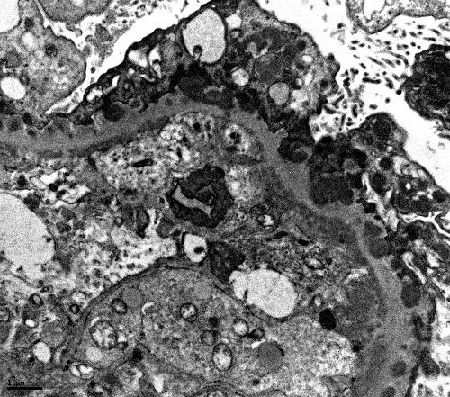
End

Previous Next
Multiple
  • Test Id: 198396adfe94861c89
  • QId: 167320
  • 35 of 96
  • Notes
  • Calculator
  • Feedback

A 25-year-old male comes to the physician complaining of flank pain and hematuria. He says the pain is constant and dull. There is no frequency, urgency or dysuria.   has a history of mental retardation and seizures. O physical exam, his blood pressure is 140/90 mm Hg, and his pulse is 80 bpm. He has multiple yellow papules on his nose and cheeks and numerous aranched skin spots on his face. A 2-3 cm hypopigmented macule is noted on the right arm. CT scan of the head was done as the patient presented with seizures. CT head w s reported normal. CT scan o the abdomen shows bilateral hypodense, fat-containing genital masses and cysts. 


What is the MOST likely diagnosis associated with these findings?

A.  Von Hippel Lindau
B.  Sturge Weber Syndrome
C. Tuberous Sclerosis
D.  Osler Weber Rendu
E.  Neurofibromatosis type 2
Correct
The correct answer is C
Tuberous Sclerosis

Explanation:
This patient’s skin lesions are consistent with sebaceous adenomas. The findings of mental retardation, sebaceous adenomas and seizures are most consistent with tuberous sclerosis. Tuberous sclerosis is associated with renal angiomyolipomas and renal cysts. Abdominal CT can diagnose these tumors because their fat density is lower than water. In patients with flank pain and hematuria, there is an increased likelihood of co-existing renal cysts. Based on the presentation and findings, this is tuberous sclerosis; the other choices are less likely, as explained below.

(Choice A) Bilateral renal cell carcinoma is associated with Von Hippel-Lindau disease. Imaging does not suggest renal cell carcinoma.
(Choice B) Leptomeningeal Angiomas are cerebral malformations commonly found in Sturge-Weber Syndrome. CT head is normal in this patient.
(Choice D) Osler-Weber-Rendu is an autosomal dominant disease associated with telangiectasias of the mucosal surfaces, as well as AVMs in the brain, GI tract and lung. Again, the clinical presentation and imaging do not suggest this diagnosis.
(Choice E) Neurofibromatosis type 2 is an autosomal dominant condition associated with acoustic neuromas, gliomas and ependymomas. The skin shows neurofibromas, but other associations favor tuberous sclerosis.

 ASSOCIATED FINDINGS IN TUBEROUS SCLEROSIS -- 
(This patient has the majority of these as bolded below)
 o      Bilateral renal angiomyolipomas (Fat-containing renal masses on CT scan)
 o      Renal Cysts
 o      Astrocytomas
 o      Cortical tubers
 o      Ash-leaf spots on skin
 o      Sebaceous adenomas on face
 o     Seizures
 o      Mental retardation

IMPORTANT TOPIC FROM RENAL BOARD POINT OF VIEW

Copyright © ABIM Exam World
Created On: 09/13/2017
Last Modified: 10/17/2026

End

Previous Next
Multiple
  • Test Id: 198396adfe94861c89
  • QId: 167322
  • 36 of 96
  • Notes
  • Calculator
  • Feedback

A 15-year-old boy is brought to the ER by his foster mother, who states that when she got home from work, she noticed he was acting very strange. He had slurred speech and seemed confused. He appeared very uncoordinated, and she was unsure whether he had fallen or hit his head. She states that he is somewhat a troubled boy but doesn’t know much about his history, as he has been in and out of the foster care system out of state. On physical exam, he is tachycardic and has tachypnoea. Pupils are dilated, but there is no nystagmus. Fundoscopic exam shows optic disc hyperemia. He is relatively uncooperative but not aggressive or hostile. When asked about suicidal thoughts, he responds only with inaudible mumbling. His foster mother left for work 10 hours prior and assumed he left for school. She is unsure when these symptoms began or what may have triggered them. P is 105/ min, BP is 140/90 mm Hg, RR is 28/min, and T is 97.1 F. Laboratory examination is as follows: 

Na   135 mEq/L                                            

K   5.0 mEq/L

CL   105 mEq/L

BUN  19 mg/dL

Cr   1.3 mg/dL         

HCO3  8 mEq/L  

Glucose  100 mg/dL         

pH   7.3          

pO2   90 mmHg

pCO2  22 mmHg

Measured serum osmolarity: 320 mmol/L

What is the next step in management?

A.  Gastric lavage
B.  N-acetylcystiene and activated charcoal
C. Fomepizole 
D. Fomepizole and Hemodialysis 
E.  Obtain serum levels of salycylate, methanol and ethylene glycol levels
Incorrect
The correct answer is D
Hemodialysis and fomepizole

Explanation: 
While this patient's case may appear to be vague, there are three strong indicators for the most likely diagnosis:
1)    This patient has metabolic acidosis with a profound Osmolar gap, which is a strong indicator of volatile alcohol related products. 
2)    Hyperemia of the optic disc (or other ocular symptoms), in addition to a profound anion gap, should make methanol toxicity a top differential diagnosis.  
3)    In addition to the serum anion gap, there is a serum Osmolar gap. (Measured Osm – Calculated Osm) where:

Calculated Osmolarity = 2(Na) + BUN/2.8 + Glu/18

***Anytime there is an osmolar gap in metabolic acidosis, your differentials can be narrowed to volatile alcohol-related substances. ***  --------------->    IMPORTANT ABIM BOARD POINT

The best treatment for methanol and ethylene glycol is to remove the toxic byproducts from circulation. The definitive therapy is Hemodialysis. Fomepizole is given to block alcohol dehydrogenase, thereby stopping the formation of toxic byproducts. However, it does not remove them from circulation; only dialysis can.

(Choice A) Gastric lavage is rarely the right answer, especially for an unknown ingestion. Caustic substances can cause further damage during gastric lavage. Esophageal perforation is potentially a fatal risk.

(Choice B) N-acetylcysteine is the treatment for acetaminophen toxicity; however, timing is important, and drug levels should be drawn first to determine the need for treatment. If ingestion occurred more than 24 hours earlier, treatment has no effect and is therefore not given.

(Choice C, D) Refer to the explanation section above.

(Choice E) When suspicion for methanol poisoning is high, treatment should not wait for diagnosis confirmation. Ocular involvement is an ominous sign, and the risk of blindness increases. In this case, the benefits outweigh the risk of treating before confirming the diagnosis.


Copyright © ABIM Exam World
Created On: 09/13/2017
Last Modified: 10/17/2026

End

Previous Next
Multiple
  • Test Id: 198396adfe94861c89
  • QId: 167339
  • 37 of 96
  • Notes
  • Calculator
  • Feedback

All of the following help predict AV Graft stenosis EXCEPT:

A. Hyperpulsatility of the AV Graft
B. Ruduced pulse augmentation
C. Increased bleeding and clots 
D. Decreased KT/V
E. Decreased Blood Flow rate
F. Surveillance of the graft
Incorrect
The correct answer is F.

Surveillance of the graft

Explanation:

Intra-graft or venous outflow stenosis – A strong pulse in the AV graft (hyperpulsatility) suggests increased resistance, as occurs with a venous stenotic lesion. Pulse intensity is directly proportional to stenosis severity. For this reason, hyperpulsatility in an AV graft can indicate impending AV graft stenosis.

Arterial stenosis – REDUCED PULSE AUGMENTATION suggests inflow stenosis due to stenosis of the arterial anastomosis or of the feeding artery. Although pulse augmentation is commonly performed, it is less sensitive for detecting inflow stenosis in AV fistulas than in AV grafts. EDD BLOOD CLOTS, DECREASED KT/V, and DECREASED BLOOD FLOW RATE are all suggestive of impending AV graft stenosis. Patients often have dilated collateral veins over the arm and chest wall (very important to examine your hemodialysis patients after they take off their shirts), suggesting central venous stenosis. The classic physical finding in a patient with significant central venous stenosis is diffuse upper extremity edema. Subcutaneous collateral veins are frequently evident over the chest. Swelling and collateral veins are caused by generalized venous hypertension of the extremity, which occurs in central lesions but is rare with peripheral lesions. 

SURVEILLANCE  does not predict AV Graft stenosis. It is not a sensitive or specific modality for accurate detection.

Copyright © ABIM Exam World
Created On: 09/14/2017
Last Modified: 10/17/2026

End

Previous Next
Multiple
  • Test Id: 198396adfe94861c89
  • QId: 167340
  • 38 of 96
  • Notes
  • Calculator
  • Feedback

A 40-year-old pleasant African man with ESRD secondary to FSGS started automated peritoneal dialysis. His prescription includes 2.5 L and 3 exchanges over 8 hours at night, with a last fill of 2 L. His urine output is 1000 mL/day. A typical ultrafiltration on the cycler is used at 1000 mL. The average drain volume during the day dwell was 1500 mL before going on the cycler at night. 

He came with complaints of lower abdominal wall edema extending to the scrotum over the past 5 days. Without any change in the dialysis prescription, his drain volume before going on the cycler dropped to 900 mL, and the ultrafiltration volume on the cycler came down to 100 mL. He reports no pain with fill or drain. 

What is the next step?


A. Chest X ray PA and lateral view 
B. Drain the fluid middle of the day to reduce the dwell time
C. PD catheter manipulation
D. Abdominal CT scan with contrast in the dialysate
E. Switch to hemodialysis 
F. Pleurodesis 
Correct
The correct answer is D. 

Abdominal CT scan with contrast in the dialysate.e

Explanation:

Typical ultrafiltration failure from peritoneal membrane failure presents gradually. More often, we see apparent ultrafiltration failure for other reasons. 

Catheter malfunction can present with problems with inflow, outflow or both. A kink in the catheter can cause problems with inflow and outflow. 

Omentum or blood clots clogging the catheter can cause outflow problems only. Fibrin clots, constipation with a loaded rectum, and a displaced PD catheter also cause outflow obstruction. X-ray KUB is very helpful in diagnosing a displaced catheter.   

Fluid leaking into the abdominal wall causes swelling in the lower abdomen extending to the scrotum or mons pubis and a decrease in drain volume. Usually, this can be treated by temporarily doing low-volume PD or transferring to HD for about 6 weeks. Leaks starting a few years after initiating PD usually do not respond to this approach and often require placement of a new PD catheter.

A patent tunica vaginalis testis will cause UNILATERAL OR BILATERAL SCROTAL SWELLING without much swelling of the abdominal wall, sometimes associated with a decrease in the drain volume. A CT scan of the abdomen with ‘IV’ contrast into the peritoneal fluid can help in diagnosing the leak into the abdominal wall and scrotal leak. A patent tunica vaginalis testis requires surgery. Generally, hemodialysis is not required after surgery, and low-volume peritoneal dialysis can be resumed. This patient presents with signs of fluid leaking into the abdominal wall. A chest X-ray is not required, so (Choice A) is wrong.

Draining during the day helps if the membrane is very permeable, with a very high D/P creatinine ratio on PET. This does not help in an abdominal wall leak of dialysate. Therefore, (Choice B) is also wrong.

PD catheter manipulation (Choice C) is wrong because this patient does not have a displaced PD catheter based on the information given.

(Choice D) is the correct answer. If the clinical presentation is very convincing, most patients are treated by reducing the dialysate volume and, if possible, keeping the abdomen dry during the day. Sometimes these patients need to be switched to hemodialysis for 4-6 weeks before PD is resumed. Hence, a temporary switch to HD would be correct, but an unqualified switch to HD is stated in answer E, which would be wrong. Therefore, (Choice E) is the wrong or less appropriate answer.

ABDOMINAL ADHESIONS cause a decrease in the drain volume as well and require surgical treatment with low-volume PD or temporary HD after that.

DIAPHRAGMATIC LEAK into the pleural space presents with cough and shortness of breath without peripheral signs of fluid overload a few weeks after starting peritoneal dialysis (not the presentation in this patient). Usually, such patients present with right-sided pleural effusion clinically and on chest X-ray (PA and lateral view. These patients can be treated with temporary transfer to pleurodesis and return to PD 4-6 weeks later. Permanent switch to hemodialysis without pleurodesis is another option.


TREATMENT OPTIONS OF VARIOUS ABDOMINAL/CHEST PROBLEMS AS MENTIONED ABOVE IN PATIENTS ON PERITONEAL DIALYSIS ARE FREQUENTLY TESTED ON BOARDS. PLEASE KNOW THE TREATMENT OPTIONS.


Copyright © ABIM Exam World
Created On: 09/14/2017
Last Modified: 10/17/2026

End

Previous Next
Multiple
  • Test Id: 198396adfe94861c89
  • QId: 167345
  • 39 of 96
  • Notes
  • Calculator
  • Feedback

Cyclosporine nephrotoxicity in a renal transplant recipient is associated with all the following renal biopsy findings EXCEPT:

(THIS PICTURE BELOW IN LOW POWER SHOWS ONE OF THE CLASSICAL FINDINGS IN CSA TOXICITY)


A. Interstitial Fibrosis  
B. Tubular atrophy 
C. Endothelial cell swelling 
D. Glomerular thrombin deposits
E. Glomerular basement membrane thickening 
F. Double contours of the GB 
Incorrect
The correct answer is D.

Glomerular thrombin deposits

Explanation:

In a patient with suspected cyclosporine nephrotoxicity, renal biopsy reveals obliterative arteriolopathy (classically in the afferent renal arterioles), suggesting primary endothelial damage and subsequent endothelial cell swelling, which may persist for months with elevated cyclosporine blood levels. This is also associated with a thickened glomerular basement membrane and a double-contour pattern. According to BANF, a thickened glomerular basement membrane and double-contour pattern most suggest chronic allograft nephropathy (CAN), also called transplant glomerulopathy. 

The other renal biopsy findings of cyclosporine nephrotoxicity include ischemic collapse or scarring of the glomeruli, vacuolization of the tubules, FSGS, and focal areas of tubular atrophy and interstitial fibrosis (producing a picture of "ZEBRA" or "STRIPED" fibrosis). These changes are seen with both low-dose and higher-dose cyclosporine therapy. However, they seem to correlate earlier with higher doses. 

(THE ABOVE PICTURE IN THE UPPER HALF SHOWS TUBULAR ATROPHY APPEARING DARK AND REDDISH ALTERNATING WITH LIGHT BLUE AREAS OF INTERSTITIAL FIBROSIS GIVING A "STRIPED" " ZEBRA" APPEARANCE)

THE PICTURE BELOW SHOWS TUBULAR ATROPHY, VACUOLIZATION OF THE TUBULES AND ISCHEMIC CHANGES:


Mild arteriolar hyalinosis at six months appears to be associated with high doses of cyclosporine and was reversible. However, after more than a year, irreversible severe arteriolar hyalinosis and glomerulosclerosis were observed, despite decreased doses and trough levels of cyclosporine.

Glomerular thrombin deposits are typically seen in patients with Lupus, antiphospholipid syndromes and other vasculitides. It is typically not seen in cyclosporine nephrotoxicity.

Copyright © ABIM Exam World
Created On: 09/14/2017
Last Modified: 10/17/2026

End

Previous Next
Multiple
  • Test Id: 198396adfe94861c89
  • QId: 167347
  • 40 of 96
  • Notes
  • Calculator
  • Feedback

A 68-year-old African American male with a known history of diabetes mellitus,  hyperlipidemia, CVD, and GERD presents to the ER with chest pain and shortness of breath. The patient says the pain is crushing and radiates to his left arm. It woke him from sleep 30 minutes ago. He took his antacids and 4 tablets of nitroglycerin, and the pain got better. His current medications include low-dose aspirin, metoprolol, glyburide, pioglitazone, lisinopril, simvastatin, and omeprazole. He recently suffered an upper respiratory infection for which he was given Levofloxacin. Vitals: BP 160/95 mm Hg, T 98.1, HR 110. He has no leg edema and no jugular venous distention. EKG shows LVH by voltage criteria with an isoelectric ST segment. Serial troponin levels are normal. A stress test showed evidence of stress-induced ischemia in the anterior leads. He is scheduled for cardiac catheterization the next day. Laboratory results are as follows:

Serum Chemistry:

Na   143 mEq/L

K   3.5 mEq/L

Cl   101 mEq/L

HCO3  22 mEq/L

BUN   35 mg/dL

Cr   3.0 mg/dL

Glucose  110 mg/dL 

What would you do to PROTECT the kidneys before doing the cardiac catheterization?

A. Dopamine IV infusion before and after the  procedure.
B. Nifedipine.
C. Isotonic sodium bicarbonate infusion before and after the procedure.
D. N Acetyl Cysteine before and after the procedure
E. Start mannitol IV before the procedure
F. Left ventriculogram quickly with coronary angiogram
Correct
The correct answer is C.

Isotonic sodium bicarbonate infusion before and after the procedure. N-acetylcysteine is also used, but its efficacy in studies is unproven.

Explanation:

Contrast nephropathy usually presents as a rise in creatinine 24-48 hours after exposure to IV radiocontrast.

Other features of CONTRAST NEPHROPATHY are:
  • Low fractional excretion of sodium (FeNa) below 1%.
  • Iodinated contrast agents can cause false-positive protein so that proteinuria may be overestimated.d
  • It is reversible most of the time.
  • UA may show muddy brown granular casts, tubular epithelial cell casts and numerous tubular epithelial casts.
Contrast-associatedted nephropathy has to be differentiated from atheroembolic disease, which presents with deterioration in kidney function days to weeks after the procedure,  embolic lesions (such as digital ischemia of the toes) or livedo reticularis, transient eosinophilia and hypocomplementemia,  protracted course with frequently little or no recovery of renal function, and kidney biopsy showing the typical biconvex, needle-shaped cl ""ts """ "ost"") within the occluded vessel, from the space occupied by the cholesterol crystal that dissolves during tissue processing, giving a "fish-out" appearance.

(Choice A) No evidence supports dopamine infusion as beneficial. In fact, it may be harmful.

(Choice B) No evidence supports that nifedipine is beneficial.

(Choice C) Studies suggest that infusion of isotonic saline or half-normal saline may be beneficial. Since alkalization may protect against free radical injury, sodium bicarbonate may be superior to isotonic saline. In patients who tolerate extra volume (i.e., those who are not volume overloaded or those who are not in heart failure), it would be reasonable to infuse isotonic sodium bicarbonate or saline.

(Choice D)Some studies suggest that oral N-acetylcysteine before and after the procedure reduces the risk of contrast nephropathy. Other studies suggest it is not beneficial. N-acetylcysteine is believed to work through its vasodilator and antioxidant effects. Because N-acetylcysteine is safe and inexpensive, many physicians would administer it prophylactically as well, even though results are conflicting. 

(Choice E) There are studies to suggest that prophylactic mannitol or furosemide is harmful.
    
(Choice F) Left ventriculogram should be avoided in patients at high risk of developing contrast nephropathy to minimize the quantity of IV contrast needed.

RISK FACTORS for contrast nephropathy are :
  • Chronic kidney disease
  • Diabetes mellitus
  • Congestive heart failure
  • Myeloma kidney
  • Hypotension

Educational Objective:

1. Identify and diagnose contrast nephropathy

2. Prevention of contrast nephropathy

Copyright © ABIM Exam World
Created On: 09/14/2017
Last Modified: 10/17/2026

End

Previous Next
Multiple
  • Test Id: 198396adfe94861c89
  • QId: 16521
  • 41 of 96
  • Notes
  • Calculator
  • Feedback

As per the JNC VIII committee recommendation, for individuals who are part of the African American population, including those with diabetes, the initial treatment should include a thiazide-type diuretic or calcium channel blocker (CCB).  

Which of the following thiazide-type diuretics is preferred as the initial antihypertensive therapy?


A. Hydrochlorthiazide 
B. Chlorthalidone 
C. Indapamide 
D. Metalazone 
E. Polythiazide 
Correct
The correct answer is B 
Chlothalidone

Explanation:

• Chlorthalidone at the same dose is approximately 1.5 to 2 times as potent as hydrochlorothiazide.

• Chlorthalidone has a longer duration of action than hydrochlorothiazide.

• There are no randomized clinical trials that directly compare outcomes in hypertensive patients treated with hydrochlorothiazide versus chlorthalidone. A multiple treatment (network) meta-analysis of nine trials including 50,946 patients was conducted in which hydrochlorothiazide and chlorthalidone were indirectly compared by evaluating their efficacy against common comparative drugs (ACE inhibitors were compared with hydrochlorothiazide in the ANBP2 trial and with chlorthalidone in the ALLHAT trial). The major findings of this meta-analysis showed that chlorthalidone significantly reduced the risk of cardiovascular events compared to hydrochlorothiazide (relative risk 0.79, 95% CI 0.72 to 0.88) and heart failure (relative risk 0.77, 95% CI 0.61 to 0.98). The authors calculated that 27 patients would need to be treated with chlorthalidone instead of hydrochlorothiazide over five years to prevent one cardiovascular event. Chlorthalidone remained superior to hydrochlorothiazide even after the meta-analysis was controlled for achieved office systolic blood pressure (relative risk for cardiovascular events 0.82, 95% CI 0.70 to 0.97). This finding may reflect chlorthalidone's longer duration of action and lower nocturnal blood pressure.

• Multiple Risk Factor Intervention Trial (MRFIT) - Men Hypertensive 2392 were treated with chlorthalidone, and 4049 were treated with hydrochlorothiazide. During six years of follow-up, cardiovascular events (defined as myocardial infarction, stroke, coronary artery bypass surgery, heart failure, left ventricular hypertrophy, peripheral artery disease, or angina) were significantly less common with chlorthalidone compared with hydrochlorothiazide (hazard ratio 0.79, 95% CI 0.68 to 0.92). Throughout the study, systolic blood pressure and LDL cholesterol levels were also lower with chlorthalidone than with hydrochlorothiazide. 

• Indapamide is a thiazide-like diuretic and has a half-life of 14-16 hours. This drug has been used in the HYVET trial alone or in combination with perindopril in the treatment of hypertension in patients more than 80 years old. The study showed a 30% reduction in stroke, a 39% reduction in the rate of death, a 21% reduction in death from any cause, a 23% reduction in CV death and a 64% reduction in the rate of heart failure. The trial showed that careful BP lowering in the very elderly is beneficial. Indapamide is not preferred over chlorthalidone.

• Metolazone - There are many studies with metolazone available. An important additional property is its effectiveness as a diuretic at lower GFR values. The duration of action is about 24 hours.
It is not preferred over chlorthalidone.

Copyright © ABIM Exam World
Created On: 09/20/2017
Last Modified: 10/17/2026

End

Previous Next
Multiple
  • Test Id: 198396adfe94861c89
  • QId: 16524
  • 42 of 96
  • Notes
  • Calculator
  • Feedback

Which of the following antihypertensive medications resulted in better blood pressure control and cardiovascular outcomes when combined with an ACE inhibitor?


A. Beta blocker 
B. Calcium channel blocker
C. Angiotensin Receptor Blocker 
D. Diuretics 
Correct
The correct answer is B
Calcium channel blocker 

Explanation:

The ACCOMPLISH trial clearly showed that an ACE inhibitor combined with a Calcium channel blocker (amlodipine) is better than an ACE inhibitor combined with a diuretic (hydrochlorothiazide). The study did not involve beta blockers or angiotensin receptor blockers. 

The ACCOMPLISH trial was the first major clinical trial addressing the issue of combination therapy in 11,000 patients who were at high cardiovascular risk (with prior CAD, diabetes, and/or CKD). The patients were randomly assigned to combination therapy with benazepril (Benazepril) combined with either amlodipine (5 mg/day) or HydrochlHydrochlorothiazideg/day).

The mean blood pressure was significantly lower in the benazepril group, and the primary endpoint was also significantly improved in this group. The treatment combination of benazepril-amlodipine also showed a similar benefit on the secondary endpoint of cardiovascular death compared with the group treated with amlodipine and hydrochlorothiazide. 

Copyright © ABIM Exam World
Created On: 09/20/2017
Last Modified: 10/17/2026

End

Previous Next
Multiple
  • Test Id: 198396adfe94861c89
  • QId: 165212
  • 43 of 96
  • Notes
  • Calculator
  • Feedback

The ACCOMPLISH trial is the first major trial addressing the issue of combination therapy in 11,506 patients who were at high cardiovascular risk. The goal blood pressure was <130/80 mm Hg in patients with diabetes or impaired renal function, and <140/90 mm Hg in patients with prior cardiovascular disease.

Which of the following combinations of blood pressure medications was the best in reducing cardiovascular events and slowing the progression of nephropathy in patients with hypertension who were at high risk for such events?

A. ACEI + Diuretics
B. ACEI + CCB 
C. ACEI + Beta-blocker 
D. CCB + Beta-blocker 
Correct
The correct answer is B
ACEI + CCB

Explanation:

• The Avoiding Cardiovascular Events through Combination Therapy in Patients Living with Systolic Hypertension (ACCOMPLISH) trial showed that initial antihypertensive therapy with benazepril plus amlodipine was superior to benazepril plus hydrochlorothiazide in reducing cardiovascular morbidity and mortality.

• ACCOMPLISH ACCOMPLISH was a double-blind, randomized trial undertaken in five countries (USA, Sweden, Norway, Denmark, and Finland). 11 506 patients with hypertension who were at high risk for cardiovascular events were randomly assigned via a central, telephone-based interactive voice response system in a 1:1 ratio to receive benazepril (20 mg) plus amlodipine (5 mg; n=5744) or benazepril (20 mg) plus hydrochlorothiazide (12.5 mg; n=5762), orally once daily. Drug doses were force-titrated for patients to attain recommended blood pressure goals.

• The primary endpoint was the composite of death from cardiovascular causes, nonfatal myocardial infarction, nonfatal stroke, hospitalization for angina, resuscitation after sudden cardiac arrest, and coronary revascularization.

• The benazepril-amlodipine combination was superior to the benazepril-hydrochlorothiazide combination in reducing cardiovascular events in patients with hypertension who were at high risk for such events.

Copyright © ABIM Exam World
Created On: 09/20/2017
Last Modified: 10/17/2026

End

Previous Next
Multiple
  • Test Id: 198396adfe94861c89
  • QId: 165215
  • 44 of 96
  • Notes
  • Calculator
  • Feedback

An 18-year-old male is brought to the renal clinic for evaluation of hypertension. There is no history of edema of the feet, puffiness of the face, hematuria, oliguria or recurrent urinary tract infections. He is an active football player, but lately he complains of weakness and muscle cramps. His blood pressure for the last 3-4 years has always been borderline high and during this visit was as noted below.

Physical examination shows: Pulse 100/min; all peripheral pulses are well felt. BP 150/100 mm Hg, Temp. 37.4 degrees C. He is pale. His growth is stunted. His cognitive function is normal, and no focal neurological deficits are noted. Other systems are unremarkable. Laboratory examination is as follows:

Hemoglobin   13.5 g/dL

Hematocrit   42%

Platelet Count   150,000 mm3

White Blood Cells  8,000 mm3


Urinalysis: 

Protein     100 mg/24 h

Glucose       None

Red Blood Cells      None

White Blood Cells     None

Leukocyte Esterase   Negative

Nitrites     Negative


Chemistry: 

BUN   13.5 mg/dL

Creatinine  0.9 mg/dL

Sodium   140 mEq/L

Potassium  5.8 mEq/L

Bicarbonate  18.5 mEq/L

Chloride   112 mEq/l

Calcium   9.2 mEq/L

Phosphorus  3.2 mg/dL

Aldosterone  5 ng/mL

PRA   <1.0 ng/dL/hr


Glucose   100 mg/dL

HbA1C    5.30%

S. Osmolality   282 mOsmol

Uric Acid   5.3 mg/dL

ESR    9.8 mm/h


ABG: 

pH   7.25

PCO2  32 mm Hg

HCO3 16 mEq/L

PO2   90 mm Hg

U: Normal-sized kidneys, no hydronephrosis 

Considering the history and the laboratory findings, which of the following is most likely the cause of hypertension in this patient?


A. Liddle's Syndrome 
B. Gordon's Syndrome 
C. Congenital adrenal hyperplasia 
D. Glucocorticoid-remediable aldosteronism 
E. Mineralocorticoid receptor activating mutation 
Correct
The correct answer is B
Gordon’s syndrome

Explanation:

Gordon’s syndrome - A young male with hypertension, hyperkalemia, hyperchloremic metabolic acidosis, normal renal function, low renin, and low aldosterone is highly characteristic of Gordon’s syndrome. This syndrome is also known as pseudohypoaldosteronism type II. Recently, it has been reported that Gordon’s syndrome is linked to chromosomes 1, 12, and 17. Hyperkalemia and hypertension in Gordon’s syndrome are caused by abnormalities in WNK kinases, proteins that are localized to the distal nephron and affect the thiazide-sensitive Na-Cl Cotransporter. Mutations in WNK4 or WNK1 Increase chloride reabsorption in the distal nephron, reducing lumen electronegativity and lowering the driving force for potassium secretion. These mutations also decrease expression of the potassium channels through which potassium enters the collecting duct. Thus, mutant WNK4 increases sodium chloride reabsorption in the distal tubule and decreases potassium secretion in the collecting duct, resulting in sodium retention, decreased potassium excretion, hypertension, hyperkalemia, and metabolic acidosis. Volume expansion suppresses renin and aldosterone. 

(Choice A) Liddle’s syndrome presents as hypertension in childhood. It is usually resistant to standard antihypertensive drugs. This is due to a gain-of-function mutation on chromosome 16 affecting the beta or gamma subunit of the ENaC channel in the collecting duct of the nephron. This results in sodium retention and volume expansion, causing hypertension and hypokalemia. Volume expansion suppresses renin and aldosterone. These patients will have hypertension, hypokalemia, metabolic alkalosis, low renin and low aldosterone. 

(Choice C) Congenital adrenal hyperplasia presents in childhood with hypertension and virilization in girls and precocious puberty in boys. The other features include primary amenorrhea, delayed sexual maturation, ambiguous genitalia, with clitoral enlargement in girls. Deficiencies of 11-beta hydroxylase (CYP11B1) and 17-alpha-hydroxylase (CYP17) lead to excess deoxycorticosterone, which causes hypertension through mineralocorticoid action. Biochemically, it is characterized by metabolic alkalosis, low renin and low aldosterone.

(Choice D) Glucocorticoid-remediable aldosteronism (GRA) is also known as familial hyperaldosteronism type I. In this disorder, mutations in the 11-beta-hydroxylase gene (CYP11B2) and the aldosterone synthase gene (CYP11B2) increase aldosterone production. Patients with GRA have ectopic aldosterone synthesis occurring in the zona fasciculata under the direct influence of ACTH rather than renin. GRA presents with hypertension, hypokalemia, metabolic alkalosis, low renin and high aldosterone. GRA resembles a primary hyperaldosteronism state with excessive urinary potassium loss. Look for a clue of family history of CVA or brain haemorrhage in the stem of the question.

(Choice E) A mineralocorticoid receptor-activating mutation is a very rare autosomal dominant disorder. The mutation lies in the mineralocorticoid receptor, causing it to be activated by steroids lacking 21–hydroxyl groups, such as progesterone and spironolactone. Both normally antagonize the receptor but have opposite effects on this mutant receptor. It usually presents as early-onset hypertension in females. It may get accelerated during pregnancy due to elevated levels of progesterone. Biochemically, it is characterized by hypokalemia, metabolic alkalosis, low renin and low aldosterone.

Educational objective:

IF YOU KNOW THESE 5 POINTS FOR THESE 5-6 SYNDROMES, YOU CAN ANSWER ALL NEPHROLOGY BOARD QUESTIONS ASKED ON THESE SYNDROMES:

Blood pressure - Normotension or hypertension
Potassium        - Hyperkalemia or hypokalemia
Acid-Base         - Acidosis or alkalosis
Renin                - Low or High
Aldosterone      - Low or High

Copyright © ABIM Exam World
Created On: 09/20/2017
Last Modified: 10/17/2026

End

Previous Next
Multiple
  • Test Id: 198396adfe94861c89
  • QId: 165230
  • 45 of 96
  • Notes
  • Calculator
  • Feedback

A 56-year-old male was brought to the emergency room with drowsiness and lethargy. He has had these symptoms for the past 2 days. He complains of a recent history of anorexia, nausea, and vomiting. He has diabetes mellitus and has been on glimepiride 1 mg daily for the last 4 years. One week ago, he had decreased vision with redness in his right eye. He was treated by his ophthalmologist with drops,s which seem to have resolved the problem. He currently takes cholecalciferol weekly for osteoporosis. On physical examination,n his pulse is 80/min, blood pressure is 140/90 mm Hg, respiratory rate is 20/min, and temperature is 97.7°F. The patient appears drowsy but shows no focal neurological deficits. Review of systems is otherwise unremarkable. Urinalysis is positive for glucose and negative for proteinuria, WBCs and RBC casts.A24-hour urinary protein collection shows proteinuria of 3.5 g/day. Further labs reveal:

Hemoglobin  8 gm%

Hct    24%

MCV   85

WBC   7800/ml

PMN   80%

Lymphocytes  20%

ESR   80 mm/hr

Na    145 mEq/L

BUN   80 mg/dL

Cr    1.8 mg/dL

CL    115 mEq/L

HCO3   25 mEq/L

Uric acid   5.8 mg/dL

Ca    14 mg/dl

PO4    2.8 mg/dL

Total Protein  7.8 gm/dL

Albumin   3.5 mg/dL.

Vitamin D  40 ng/ml

PTH   10 pg/ml

Which of the following is most likely the cause of his hypercalcemia?


A.  Vitamin D toxicity
B. Primary hyperparathyroidism
C.  Multiple myeloma.
D. Sarcoidosis
Correct
The correct answer is C.

Multiple myeloma

Explanation:

Multiple myeloma results from the proliferation of plasma cells that produce a monoclonal immunoglobulin.

The diagnosis of multiple myeloma is supported by the presence of many of the following factors:
  • Advanced Age
  • Backache and Bone pain
  • Anemia
  • High ESR
  • Urine protein negative by dipstick and positive by other tests is characteristic of light chain proteinuria.
  • High Total protein
  • Low anion gap
  • Hypercalcemia and hyperuricemia favor the diagnosis of multiple myeloma.

(Choice A) This patient has received Vitamin D treatment. Vitamin D toxicity can give rise to hypercalcemia. Serum vitamin D3 levels will be high. PTH will be low. Patient’s vitamin D level of 40ng/ml (normal range is 30-100ng/ml) does not favor Vitamin D toxicity.

(Choice B) Primary hyperparathyroidism is the commonest cause of hypercalcemia among outpatients. It is characterized by elevated PTH. The patient’s PTH level of 10 pg/ml rules out primary hyperparathyroidism.

(Choice D) Sarcoidosis is a multisystem granulomatous disorder of unknown cause. Hypercalcemia and hypercalciuria in sarcoidosis are associated with high vitamin D levels. This patient has low vitamin D levels; therefore, it is unlikely to be the cause. There is also no history or findings to suggest sarcoidosis.

Copyright © ABIM Exam World
Created On: 09/20/2017
Last Modified: 10/17/2026

End

Previous Next
Multiple
  • Test Id: 198396adfe94861c89
  • QId: 165233
  • 46 of 96
  • Notes
  • Calculator
  • Feedback

A 25 year-old female is referred by her primary care provider for evaluation of hypertension and hypokalemia. The primary care provider has already started her on oral potassium, despite therapy her Potassium being 2.8 meq/L. Her blood pressure despite treatment with amlodipine and Lisinopril 154/96 mm of Hg. There is no renal bruit. Systemic and fundus examinations are normal. Her mother was also diagnosed with hypertension at an early age. Her brother died of a cerebrovascular accident 2 years ago. Laboratory findings are as follows:

Na   140 

Potassium 2.8 

Chloride   100 

HCO3    26

BUN   15 

Creatinine  0.8 

Glucose    110

TSH and Cortisol are normal

ACTH    elevated

Renin   0.7 (Low)

Aldosterone  48 (elevated)


Urinalysis:

Sodium   240 mEq/D

Potassium  98 mEq/D

Urinary 18-OH Cortisol and 18-oxocortisol are elevated.

The most appropriate treatment for this patient is:

A. Steroids
B. Spironolactone 
C. Steroids + Spironolactone
D. Amiloride 
Correct
The correct answer is C

Steroids + Spironolactone

Explanation:

This patient has (GRA) – Glucocorticoid Remediable Aldosteronism. These are typically young patients presenting with hypertension and hypokalemia. There is strong family history of early onset hypertension along with a history of fatal CVA or hemorrhagic strokes. These patients typically have low Renin and high Aldosterone mimicking a state of primary hyperaldosteronism. Because of this they have high urinary potassium losses as manifested in this patient. (Urinary potassium of more than 40mEq/D is considered increased losses). Increased ACTH and 18-OH cortisol further support the diagnosis of GRA.

Steroids alone are used in the treatment of Pregnancy associated mineralocorticoid excess and congenital adrenal hyperplasia. Steroids alone in this patient will suppress ACTH but this patient also needs an aldosterone antagonist for the hyperldosterone state and to reduce urinary potassium losses. Hence, the right answer is steroids plus spironolactone.

Spironolactone alone is not sufficient, as steroids are needed to suppress the increased ACTH state. 

Amiloride blocks the EnaC channel in Liddle’s syndrome.

PLEASE NOTE THAT "GRA" IS A FREQUENTLY ASKED QUESTION IN NEPHROLOGY BOARD EXAMS

PLEASE REVIEW GLUCOCORTICOID REMEDIABLE ALDOSTERONISM WELL. 

Copyright © ABIM Exam World
Created On: 09/20/2017
Last Modified: 08/06/2018

End

Previous Next
Multiple
  • Test Id: 198396adfe94861c89
  • QId: 165242
  • 47 of 96
  • Notes
  • Calculator
  • Feedback

A 60-year-old with recently diagnosed colon cancer and diabetes presents with bilateral pedal edema; BP is 120/80 mm Hg. Urinalysis showed  4+ protein, no RBCs or WBCs, and 8-10 Hyaline casts. His BUN is 20, Creatinine is 1 mg/dL, and albumin is 2 grams/dL. A 24-hour urine collection showed 10 grams of protein. The patient undergoes a kidney biopsy. The EM is shown below :


What is the most likely diagnosis?

A.  Crescentic GN
B.  IgA nephropathy
C.  Minimal change disease
D.  Focal segmental glomeruloscerosis
E.  Membranous nephropathy
F. Diabetic nephropathy
Correct

The correct answer is E

Membranous Nephropathy.

Electron microscopy shows subepithelial electron-dense deposits, as classically seen in membranous nephropathy. If in the question there is a suggestion of colon, breast, or lung cancer, then the glomerulopathy is usually membranous. Then look for other histopathological findings that confirm the diagnosis: subepithelial electron-dense deposits.

Explanation:

BOARD POINT - FAMILIARIZE YOURSELF WITH THESE ASSOCIATIONS :

1. Solid cancers (colon, breast, lung, renal) plus proteinuria = Membranous nephropathy

2. Hodgkin's lymphoma plus proteinuria = Minimal change disease

3. HIV plus proteinuria = Focal segment glomerulosclerosis FSGS

4. Pamidronate plus protenuria = FSGS (rare)

5. Myeloma, no albuminuria on dipstick, but proteinuria on protein/creatinine ratio or 24-hour urine: Light chain nephropathy

6. Myeloma with nonspecific proteinuria (dipstick, urine protein/creatinine ratio and 24-hour urine): Light chain nephropathy or amyloidosis.


BOARD POINT - FAMILIARIZE YOURSELF WITH THESE HISTOPATHOLOGY ASSOCIATIONS FOR VARIOUS GLOMERULAR DISEASES


Copyright © ABIM Exam World
Created On: 09/20/2017
Last Modified: 10/17/2026

End

Previous Next
Multiple
  • Test Id: 198396adfe94861c89
  • QId: 165243
  • 48 of 96
  • Notes
  • Calculator
  • Feedback

You are the nephrologist on call. The  ER calls you for an 18-year-old female who is complaining of vomiting and diarrhea. Her serum sodium is 116 mEq/L and Serum potassium is 5.9 mEq/L. On physical examination, the patient is drowsy;p; pulse is 126/min, BP is 90/60 mm Hg, and RR is 32/min. Her chest is clear. Her heart sounds are normal, with no murmurs. The patient is drowsy but arousable, with no focal neurological deficit. Laboratory findings are as follows:

Hb    16 gm/dl 

WBC   12,800/cmm 

Polymorph  46% 

Lymphocytes  16% 

Eosinophils  4%

Monocytes  4%

Platelets   2,40,000/cmm.

CL    70 mEq/L

BUN  10 mg/dl

Creatinine  0.5 mg/dl

Na    116 mEq/L

K    5.8 mEq/L


Urinalysis:

pH    6.4

Protein  trace

Glucose  absent

Microscopic: occasional WBCs & RBCs

Urinary Na  90 mEq/L

Urinary K         20 mEq/L

ABG    

PH                   7.32

PCO2   36 

HCO3   20 mEq/L

PaO2   92

O2 saturation  98%

S. Cortisol  6.00 mg/dl

TSH   3.5 IU/m (Normal 0-5 IU/m ).

Both plasma Renin and Aldosterone are high. 

Which of the following conditions is most likely with these findings?


A. Type 4 RTA 
B. Pseudo-hypo-aldosteronism Type 1 
C. Gordon's syndrome
D.  Diarrhea
Incorrect
The correct answer is B.
 Pseudo-hypoaldosteronism type 1

Explanation:

This patient has hyponatremia with high urinary sodium, high serum potassium, low urinary potassium, and normal adrenal function. The serum aldosterone level is high, suggesting target-organ resistance to aldosterone. These findings are characteristic of pseudohypoaldosteronism type 1 (PHA type 1)

PHA type 1 is a rare hereditary disorder characterized by generalized resistance to aldosterone action. It presents with 
1. Salt wasting 
2. Hypovolemia 
3. Normotension
4. Metabolic acidosis 
5. Hyperkalemia
6. High Renin and Aldosterone.  

NEPHROLOGY BOARD EXAM TAKERS REMEMBER THESE 6 POINTS FOR PHA TYPE 1

These are two different modes of inheritance:
1] Autosomal recessive.
2] Autosomal dominant.

Autosomal recessive affects the epithelial sodium channel and other target organs, such as the kidney, colon, and sweat gland. It causes downregulation of sodium channels and decreased sodium transport.

The autosomal dominant or sporadic form is due to heterozygous mutations in the NR3C2 gene, which codes for the mineralocorticoid receptor. This is a milder form than autosomal recessive disease, which affects only the kidney. The disease often improves with age.

Treatment consists of a high-salt diet. This prevents volume depletion and,d by enhancing sodium delivery to the distal tubules, increases potassium excretion, thereby lowering serum potassium. High-dose fludrocortisone (1 to 2 mg/d), or carbenoxolone, is indicated if high-salt intake is ineffective or not tolerated.

(Choice A) Type IV RTA will have low aldosterone and low renin levels. Therefore, that is not the likely answer.

(Choice D) Diarrhea will have metabolic acidosis with hypokalemia, and Net urine charge will be negative. This patient has positive Net urine charge with hyperkalemia, which rules out diarrhea as a cause.

(Choice C)  = PHA TYPE 2 (Pseudohypoaldosteronism type 2) OR = Gordon’s syndrome is characterized by:
 1. Hypertension 
 2. Hyperkalaemia 
 3. Metabolic acidosis
 4. Low plasma Renin and Aldosterone. This is due to mutations in WNK kinases 1 and 4. These mutations increase chloride reabsorption and sodium retention, resulting in hypertension.

NEPHROLOGY BOARD EXAM TAKERS REMEMBER THESE 4 POINTS FOR PHA TYPE 2 or GGORDON'SSYNDROME

IF YOU KNOW THESE 10 POINTS OF PSEUDOHYPOALDOSTERONISM TYPE 1 & 2, THEN YOU CAN ANSWER ALL QUESTIONS ON PHA 1, 2 AND GORDON'S SYNDROME WHICH ARE GOING TO BE ASKED FOR SURE ON THE BOARDS.

Copyright © ABIM Exam World
Created On: 09/20/2017
Last Modified: 10/17/2026

End

Previous Next
Multiple
  • Test Id: 198396adfe94861c89
  • QId: 165246
  • 49 of 96
  • Notes
  • Calculator
  • Feedback

A 32-year-old male is brought to the renal clinic with a history of hematuria, oedema of the feet, and puffiness of the face. He reports fever and sore throat a week ago. He also complains of breathlessness on exertion and oliguria. Physical examination shows: Pulse 100/min, BP 150/100 mm Hg, and Temp. 37.4 C. He is pale. He has facial puffiness and oedema of the feet. Systemic examination-unremarkable. Laboratory examination is as follows:

Hb   10.5 g/d

Hct   34%

Platelet 250,000 mm3

WBC  8,000 mm3

Differential count: P 80% L 12% E 6% M 2%

ESR  9.8 mm/h


Urinalysis:

Protein   3000 mg/24 h

Glucose   None

RBC   50-60/hpf Dysmorphic

WBC   occasional

Leukocyte Esterase Negative

Nitrite Negative

BUN   40 mg/dL

Creatinine  3.9 mg/dL

Sodium   140 mEq/L

Potassium  4.2 mEq/L

Bicarbonate  25.5 mEq/L

S. protein  5.5 g/dl

S. Albumin  2.5 g/dl

Calcium   9.2 mEq/L

Phosphorus  3.2 mg/dL

Glucose   100 mg/dL

Uric Acid   5.3 mg/dL

C 3    Low

C4     normal

HBsAg /HIV   Neg

ANA    Neg

Kidney Biopsy: Shows enlarged Glomeruli, lobular accentuation, mesangial hypercellularity, endo-capillary proliferation and double contour along the capillary wall. IF shows bright C3 in the mesangium and capillary wall with absent immunoglobulin staining. 

Electron Microscopy: Suggestive of dense deposits.

What is the BEST treatment option for this patient?


A. Plasma exchange + Rituximab 
B. Rituximab 
C. Eculuzimab
D. Cyclophosphamide + Steroids 
Correct
The correct answer is C.

Eculuzimab

Explanation:

This patient has a nephritic-nephrotic picture with low C3. The Kidney biopsy, along with the clinical presentation, suggests MPGN. Negative immunoglobulins, along with positive C3 staining, narrow it down to DDD (DENSE DEPOSIT DISEASE) or C3-GN (C3 glomerulonephritis)

DDD, or dense deposit disease, is best treated with Eculizumab. 
Rituximab is not useful in DDD or C3-GN. 

ECULUZUMAB is useful in:
1. DDD
2. Atypical HUS – used along with plasma exchange. If using Eculizumab, give the meningococcal vaccine or give penicillin till the vaccine becomes effective.

However, (additional information nnot aboutthis question)

RITUXIMAB: is useful in:
1. ANCA vasculitis (can be used in induction or relapse – RAVE TRIAL)
2. Wegener's
3. HCV cryoglobulinemia
TTP

***** TREATMENT OPTIONS IN GLOMERULONEPHRITIS WITH RITUXIMAB AND ECULIZUMAB ARE FREQUENTLY TESTED CONCEPTS IN NEPHROLOGY BOARD EXAMS. PLEASE REVIEW THESE IN DETAIL.*****


Copyright © ABIM Exam World
Created On: 09/20/2017
Last Modified: 10/17/2026

End

Previous Next
Multiple
  • Test Id: 198396adfe94861c89
  • QId: 165247
  • 50 of 96
  • Notes
  • Calculator
  • Feedback

A 36-year-old female was diagnosed with membranous nephropathy secondary to SLE. Her 24-hour protein excretion was 7.5 gms/day. Her serum creatinine was 0.9mg/dl. She was started on 500 mg of cyclophosphamide IV every 15 days (Euro-Lupus) and prednisolone 1 mg/kg orally per day. After 3 months of therapy, she presented with decreased urine output, puffiness of the face, and oedema of the feet. On physical examination, her temperature was 37°C, blood pressure was 160/100 mm Hg, pulse was 90/min, and respiratory rate was 20/min. She is anemic, with facial puffiness and oedema of the feet. On systemic examination, air entry was decreased at both lung bases, and heart sounds were distant and feeble. Chest X-Ray reveals bilateral pleural effusions. Echocardiogram reveals mild to moderate pericardial effusion. Laboratory examination is as follows: 

Hemoglobin   10.0 g/dL

Hematocrit   34%

Platelet Count   150,000 mm3

WBC    8,000 mm3

Differential count: P  80% L 12% E 6% M 2%

ESR    50.8 mm/h


Urinalysis: 

Protein  1450 mg/24 h

Glucose  None

RBCs  70-80/HPF dysmorphic

WBCs  5-8/HPF

Leukocyte Esterase Negative

Nitrites  Negative

 BUN   35 mg/dL

Creatinine  3.9 mg/dL

Sodium   140 mEq/L

Potassium  5.2 mEq/L

Bicarbonate  15.5 mEq/L

Calcium   9.2 mEq/L

Phosphorus  5.6 mg/dL

Glucose   100 mg/dL

Uric Acid   5.3 mg/dL

C3 & C4 decreased 

ANA   positive

dsDNA   positive

Repeat biopsy shows:


Which of the following is the most appropriate therapy for her current condition?


A. Mycophenolate and steroid
B. Mycophenolate, tacrolimus and steroids 
C. Rituximab 
D. I.V Immunoglobulins 
E. Plasma exchange  
Correct
The correct answer is B.

Mycophenolate, tacrolimus and steroids 

Explanation: 

This patient has membranous lupus being treated with the Euro-Lupus regimen. Urine activity, plus positive ANA and dsDNA and low C3 and C4, suggests disease activity. The biopsy shows active lupus nephritis combined with features of class IV & V diffuse proliferative GN. Patients with both DPGN and membranous lesions are more likely to be resistant to standard induction regimens with cyclophosphamide or MMF with steroids. In these patients, combined treatment with MMF and tacrolimus is recommended. This was suggested by Bao H. et al. in a small, short-term prospective trial in which 40 patients with diffuse proliferative plus membranous lupus nephritis were randomly assigned to induction therapy with MMF (0.75g to 1g/d) and tacrolimus (3-4 mg/d) or intravenous cyclophosphamide alone. All patients received steroids. At nine months, there was a significantly higher rate of complete remission in patients treated with MMF and tacrolimus as compared to cyclophosphamide (65% versus 15%).

(Choice A) Mycophenolate and steroids can be used in patients who have received cyclophosphamide and are resistant to it.

(Choice C and D) Rituximab and I.V. Immunoglobulins can be used in patients with lupus nephritis who have failed to respond to cyclophosphamide, MMF and steroids.

(Choice E) Plasma exchange is recommended in patients with SLE and TTP. 

KDIGO Clinical Practice Guidelines for Glomerulonephritis recommend the following treatment for resistant lupus nephritis:

Treatment of resistant disease 12.9.1: In patients with worsening S. Cr and/or proteinuria after completing one of the initial treatment regimens, consider performing a repeat kidney biopsy to distinguish active LN from scarring. (Not Graded) 12.9.2: Treat patients with worsening S. Cr and/or proteinuria who continue to have active LN on biopsy with one of the alternative initial treatment regimens. If the patient has received cyclophosphamide, use MM, F, F, and if the patient has received MMF, use cyclophosphamide (Not Graded) 12.9.3 

KDIGO suggests that non-responders who have failed more than one of the recommended initial regimens (cyclophosphamide, MM, F, or CNIs) may be considered for treatment with rituximab, IV Immunoglobulins, or CNIs. (2D).


Copyright © ABIM Exam World
Created On: 09/20/2017
Last Modified: 10/17/2026

End

Previous Next
Multiple
  • Test Id: 198396adfe94861c89
  • QId: 165256
  • 51 of 96
  • Notes
  • Calculator
  • Feedback

A 30-year-old man comes to your office with a painful rash on his neck. He has a fever and malaise. He has a history of  IV. He is currently taking Tenofovir, emtricitabine, and indinavir. The rash is suggestive of Herpes Zoster rash :

Physical examination reveals no oral cavity lesions. His current CD4 count is 250/ m3. His chemistry is normal. He is started on an intravenous medication for his rash. Two days later, his chemistry is as follows:

Na    135 mEq/L

K     4.5 mEq/L                  

CL    100 mEq/L

HCO3   24 mEq/L                  

BUN  21 mg/dL                       

Cr   2.0 mEq/L

Glucose  95 mg/dL

Calcium   9.4 mg/dl

Urinalysis shows needle-shaped crystals in the sediment.

Which of the following is most likely the cause of his renal problem?


A.  Indinavir
B.  Tumor lysis syndrome
C.  Antifreeze ingestion
D.  Acyclovir
E.  IV TMP/SMX
Correct
The correct answer is D.

Acyclovir

Explanation:

This patient has a characteristic herpes zoster lesion on his neck. The vesicles are 2-3 mm in size with an erythematous base. They are in different stages of development. Herpes zoster is commonly seen in the elderly and immunocompromised. This patient is 30 years old and immunocompromised (HIV positive with AIDS). The treatment of choice for herpes zoster in an immunocompromised patient is IV acyclovir. High-dose acyclovir is one of the causes of crystal-induced nephropathy. The crystals are needle-shaped.

Indinavir (Choice A), a protease inhibitor, is a common cause of nephrolithiasis in HIV patients. Patients would often present with flank pain..Urinalysis would show hematuria and needle-shaped crystals. This patient, however, had normal serum chemistry on presentation. Following two days of IV medication (acyclovir), his serum chemistry showed elevated creatinine, suggesting acyclovir as the most likely cause.

Tumor lysis syndrome (Choice B) occurs in the setting of chemotherapy for lymphoma. It leads to the formation of needle-shaped uric acid crystals. This patient has no such presentation.

Patients with antifreeze ingestion (Choice C) present with metabolic acidosis with an elevated anion gap. Initial urinalysis shows envelope-shaped oxalate crystals.

Trimethoprim-sulfamethoxazole (TMP/SMX) (Choice E) has sulfonamide in it. Sulfonamide also leads to crystal-induced nephropathy. The crystals are often dumbbell-shaped.

Educational Objective:

Drugs causing Crystal-induced nephropathy:

ACYCLOVIR --- NNeedle-shapedcrystals

INDINAVIR --- Needle-shaped crystals

SULPHONAMIDE --- Dumbbell-shaped crystals

CALCIUM OXALATE anantifreeze-- Envelope-shaped crystals

CALCIUM PO4----Coffin-shapedd crystals

URIC ACID --- Hexagonal crystals

CYSTEINE --- Hexagonal crystals


Copyright © ABIM Exam World
Created On: 09/20/2017
Last Modified: 10/17/2026

End

Previous Next
Multiple
  • Test Id: 198396adfe94861c89
  • QId: 165258
  • 52 of 96
  • Notes
  • Calculator
  • Feedback

 In patients with CKD and ESRD, Vitamin D appears to protect against cardiovascular events by controlling secondary hyperparathyroidism. The OPERA trial evaluated the effect of the Vitamin D analog paricalcitol in CKD stages 3-5 with left ventricular hypertrophy. The study observed the effect on left ventricular mass and function.

Which of the following statements is true about the effect of paricalcitol in CKD and ESRD patients?

A. Paricalcitol treatment reduced LV mass
B. Paricalcitol treatment preserved LV ejection fraction
C. Paricalcitol treatment improved pulse wave velocity and arterial stiffness
D. Paricalcitol treatment improved blood pressure control
E. Paricalcitol treatment significantly reduced PTH and alkaline phosphatase 
Correct
The correct answer is E

Paricalcitol treatment significantly reduced PTH and alkaline phosphatase.

Explanation:

Vitamin D Rx DOES NOT reduce LV mass OR LV ejection fraction OR arterial stiffness.s
Vitamin D seems to protect against cardiovascular disease, but its effect on patient outcomes in CKD is controversial.
The 
OPERA trial was a prospective, double-masked, randomized, placebo-controlled trial to determine whether oral activated vitamin D reduces left ventricular (LV) mass in patients with stages 3-5 CKD and LV hypertrophy.
Subjects with echocardiographic criteria of LV hypertrophy were randomly assigned to receive either oral paricalcitol (1 ?g) once daily (n=30) or matching placebo (n=30) for 52 weeks.

The primary endpoint was change in LV mass index over 52 weeks, which was measured by cardiac magnetic resonance imaging. Secondary end points included changes in LV volume, echocardiographic measures of systolic and diastolic function, biochemical parameters of mineral bone disease, and measures of renal function.
Change in LV mass index did not differ significantly between groups (median [interquartile range], -2.59 [-6.13 to 0.32] g/m(2) with paricalcitol versus -4.85 [-9.89 to 1.10] g/m(2) with placebo).
Changes in LV volume, ejection fraction, tissue Doppler-derived early diastolic and systolic mitral annular velocities, and the ratio of early mitral inflow velocity to early diastolic mitral annular velocity did not differ between groups.

Paricalcitol treatment significantly reduced intact parathyroid hormone (P<0.001) and alkaline phosphatase (P=0.001) levels, as well as cardiovascular-related hospitalizations, compared with placebo.

52 weeks of treatment with oral paricalcitol (1 ?g one time daily) significantly improved secondary hyperparathyroidism but did not alter measures of LV structure and function in patients with severe CKD and ESRD.

OPERA and PRIMO are two recent randomized clinical trials in patients with CKD that evaluated paricalcitol for slowing ventricular mass progression. Both of these trials showed negative results.

Copyright © ABIM Exam World
Created On: 09/20/2017
Last Modified: 10/17/2026

End

Previous Next
Multiple
  • Test Id: 198396adfe94861c89
  • QId: 165260
  • 53 of 96
  • Notes
  • Calculator
  • Feedback

You are rounding on your patients in the dialysis unit and see a 65-year-old gentleman with ESRD due to chronic interstitial disease. He also has a history of diet-controlled diabetes mellitus and hypertension. His other past medical history is significant for dyslipidemia, coronary artery disease, hypothyroidism, gout and depression. He has been hospitalized in the recent past for swelling and pain of his right great toe. He was seen by the foot doctor, a scan was done, and eventually the great toe had to be amputated. He has been on hemodialysis 3 times a week. His weekly Kt/V is 1.9. You are conducting the monthly blood work review for this patient. You note that his hemoglobin has been persistently low over the past few monthly blood draws. He is currently on 100 mcg of Darbepoetin weekly on dial sis. On enquiry, there is no history of blood loss in the form of hematemesis, melena, hematochezia or hemoptysis. Activee medication list includes Losartan, Atorvastatin, Calcitriol, multivitamin supplements, paroxetine, allopurinol, aspirin.

His pertinent blood work is as follows:

Test

Result

WBC

4500 cells /cumm

Hemoglobin

8.2 g/dL

Platelet count

450 thousand /cumm

Reticulocyte count

Normal

Iron

55 (range 50-150)

Total iron-binding capacity

250 g/dl (range 250-310)

Transferrin saturation

20%

Ferritin

1400 ng/ml (range 20-235)

Haptoglobin

400 mg/dl (range 83-267)

Lactate dehydrogenase

240 U/L (range 80-225)

Total bilirubin

1.0 mg/dl

Folate

7 ng/ml (range 1.8-9.0)

B12

500 pg/ml (range 200-800)


Peripheral blood smear.

Normal RBC morphology, few burr cells.


Which of the following is true about this patient’s anemia?


Omitted

Correct Answer: Option C: This patient has a chronic inflammatory state that contributes to his anemia.


Explanation:

10-15% of patients receiving erythrocyte-stimulating agents ESAsAs) develop resistance. Here are multiple reasons why ESRD patients develop resistance.


ESA resistance occurs due to the following reasons:

  1. Iron deficiency.

  2. Chronic inflammation.

  3. Under-dialysis.

  4. Hemolysis.

  5. Folate and B12 deficiency.

  6. Chronic blood loss.

  7. Anti-EPO antibodies.

  8. Pure red cell aplasia.

  9. Failed chronic renal allograft.

  10. ACEI/ARB.

  11. Aluminum overload.

  12. Hyperparathyroidism.

  13. Hematological disorders or malignancy.


Option A: Incorrect option. SRD is associated with erythropoietin deficiency. The patient has already been initiated on ESA. There is no point in measuring EPO levels. There is no evidence that measuring EPO levels is part of anemia management in CKD.

Option B: Incorrect option. SRD is an inflamed state. In an inflammatory milieu, hepcidin production increases. The hepatic iron-regulatory hormone Hepcidin and its receptor, the cellular iron exporter Ferroportin, form a feedback-regulated mechanism that maintains adequate plasma iron-transferrin concentrations for erythropoiesis and other functions, ensures sufficient iron stores, and prevents iron toxicity. In chronic kidney disease, inflammation and impaired renal clearance increase plasma hepcidin, which inhibits duodenal iron absorption and sequesters iron in macrophages. These hepcidin effects can cause systemic iron deficiency, reduced iron availability for erythropoiesis, and resistance to endogenous and exogenous erythropoietin.

Choice C: Correct option. Refer to the explanation for option B. He had pain and swelling of his right great toe; a foot doctor saw him, a bone scan was done and subsequently, ly the amputation. All findings suggest an infective etiology, probably osteomyelitis. There is a temporal relationship between the patient's anemia and an underlying chronic inflammatory state.

The high ferritin also suggests an inflammatory state.

Choice D: Incorrect option. Pure red cell aplasia, a form of severe ESA hypo-responsiveness mediated by anti-erythropoietin antibodies, was first reported with certain formulations of Epoetin alfa but has now been reported with all commercially available forms of ESA. This syndrome presents with rapid-onset severe anemia (hemoglobin <7 gg/dL, severe reticulocytopenia (reticulocyte count <10,000/l L, and marked elevations in serum ferritin (>1000 ng/mmL and transferrin saturation (>70%) due to low iron utilization. Pure red cell aplasia is unlikely given the absence of characteristic laboratory findings. Moreover, the patient did not receive Epoetin alfa.


Choice E: Incorrect option.Under-dialysis leads to anemia through the same mechanism mentioned earlier in option B. Under-dialysis worsens the uremic milieu, which in turn leads to an inflammatory state. The patient in this clinical vignette has been dialysed appropriatel   is weekly Kt/V is 1.9, which is above the target goal of 1.7

Copyright © ABIM Exam World
Created On: 10/22/2018
Last Modified: 10/16/2026

End

Previous Next
Multiple
  • Test Id: 198396adfe94861c89
  • QId: 165261
  • 54 of 96
  • Notes
  • Calculator
  • Feedback

50-year-old female patient whose group B is being evaluated for kidney transplant surgery. She had ESRD secondary to analgesic nephropathy and has been on hemodialysis for the last 5 years. She has had multiple sensitization events in the form of 3 pregnancies and several blood transfusions. Her current calculated PRA against class I antigen is 97% and against class II antigen is 99%. She has been enrolled in the national highly sensitized recipient program.

Her husband, who is blood group matched, came forward as a potential kidney donor, but she had a positive flowB andT-cell crossmatch against him. Single antigen bead assay demonstrated donor-specific antibodies against class II across DQB*15 and DPB*14. This transplant did not materialize as the patient declined the desensitization protocol. Now her younger brother comes forward as a potential donor. He is blood group A, and the flow B and T-cell crossmatch is negative, with no demonstrable donor-specific antibodies against this donor. The patient wants to know more about ABO-incompatible transplant.


Which of the following statements about the ABO incompatible transplant is correct?


A. Three-year graft survival is inferior to blood group compatible transplants.
B. The infectious and bleeding complications post ABOI kidney transplant are the same as blood matched kidney transplant.
C. All patients undergoing ABOI transplant need to undergo desensitization using IVIg, Plasma exchange, Rituximab irrespective of their donor/recipient pair Anti ABO titers for optimal outcomes.
D. C4d staining on protocol biopsies is common feature and does not necessarily mean an antibody mediated rejection process in the absence of allograft dysfunction.
Incorrect

Correct answer: Option D: C4d staining on protocol biopsies is a common feature and does not necessarily mean an antibody-mediated rejection process.


Explanation:


Choice A: Three-year graft survival is inferior to blood group-compatible transplants is incorrect. A comprehensive database analysis of 1420 ABOI living donor (LD) kidney transplants performed in 101 centers from 2005 to 2012 compared graft and patient survival with a matched cohort of ABO-compatible transplant recipients. Three-year graft and patient survival were ultimately identical. 1


Choice B: The infectious and bleeding complications post ABOI kidney transplant are the same as in blood-matched kidney transplant is also incorrect. Using USRDS and Medicare data from 2000–2007, 119 ABOI (non-A2 donor) transplant recipients were identified. Compared with ABO-compatible recipients, the risks of infectious and hemorrhagic complications were significantly higher, with a 2.2-fold higher risk of pneumonia, a 3.5-fold higher risk of wound infections, a 56% higher risk of pyelonephritis, and a nearly 2- fold higher risk of hemorrhage 2


Choice C: All patients undergoing ABOI transplant need to undergo desensitization using IVIg, Plasma exchange, Rituximab irrespective of their donor/recipient pair. Anti-ABO titers for optimal outcomes is also an incorrect answer. Historically, ABOI transplantation has been successful when performed after desensitization with plasmapheresis, intravenous Ig (IVIG), rituximab, and/or splenectomy to achieve ABO IgG antibody titersof  1:4. A recent publication demonstrated that these intensified treatments might not be necessary in donor/recipient pairs who have low-to-moderate titer ABO incompatibility 3


Choice D: C4d staining on protocol biopsies is a common feature and does not necessarily mean an antibody-mediated rejection process; in the absence of allograft dysfunction, this is the correct answer. C4d staining is not an uncommon feature seen in the protocol biopsies done in ABOI kidney transplant recipients. In the absence of allograft dysfunction, C4d staining has no clinical relevance and is part of graft accommodation.

Copyright © ABIM Exam World
Created On: 10/30/2018
Last Modified: 10/16/2026

End

Previous Next
Multiple
  • Test Id: 198396adfe94861c89
  • QId: 165262
  • 55 of 96
  • Notes
  • Calculator
  • Feedback

A 28-year-old Caucasian gentleman comes to the clinic for a follow-up visit. He has been found to have type 1 diabetes mellitus since the age of 12 years. His cousin has the same disease. He reports no symptoms and uses an insulin pump with insulin aspart. He monitors blood sugar with a flash glucose monitor and uses carbohydrate counting for boluses. He reports infrequent hypoglycemic episodes, particularly 2 hours post-lunch, but manages them by himself. He exercises at least 60 minutes per day. His vitals show BP of 118/6 mmHg. His BMI i 23.2. Physical examination is unremarkable.


His laboratory investigations are reported as follows.


Characteristic

Value

Hemoglobin

14.2 gm/L 

WBC count

6.8 X 10³/cubic mm

Platelet count

241 X 103/cubic mm

Segmented Neutrophils 

Lymphocytes

Monocytes

Band neutrophils 

Eosinophils

Basophils

60%

36%

2%

0%

2%

0%

Sr Sodium

139 mEq/L

Sr Potassium

4.4 mEq/L

Sr Creatinine

0.6 mg/dL

eGFR using CKD-EPI

136.8 ml/min/1.73m2

Sr Bicarbonate

24 mEq/L

Sr Chloride

101 mEq/L

Total Bilirubin

1.0 mg /dL

AST

16 U/L

ALT

18 U/L

Sr Albumin

4.0 g/dL

HBA1C

7.9%

Sr Calcium

10 mg/dL

Urine dipstick

pH- 5.4

Albumin-nil

 no blood

 no WBCs

24-hour urinary protein 

76 milligrams/day


What is the MOST LIKELY incorrect statement regarding the hyperfiltration stage of Diabetic Kidney Disease in this patient?

A. Renal hyperfiltration is usually diagnosed when the GFR is more than 120 ml/min,which corresponds to a renal function that exceeds two standard deviation above mean GFR.
B. Renal hyper filtration usually precedes microalbuminuria in type 1 diabetes mellitus.
C. Renal hyper filtration is considered as a risk factor for future progression to chronic kidney disease (CKD) and end stage renal disease (ESRD) in type 1 DM.
D. eGFR equations like MDRD equation can be used predict hyper filtration.
Incorrect

The Correct Answer is Option D: eGFR equations like the MDRD equation can be used pto redict hhyperfiltration


Explanation:


Supraphysiologic elevation in GFR is observed early in the natural history of type 1 and type 2 diabetes mellitus and is due to glomerular hyperfiltration. Pathogenesis of hyperfiltration in diabetes is complex, with a prominent role for hyperglycemia and altered insulin levels, especially in early diabetes and prediabetes. Dilatation of the afferent (pre-capillary) glomerular arteriole plays an important role in the hyperfiltration response, by raising both the intra-glomerular pressure and renal blood flow.


Direct measurement of GFR is usually required to detect hyperfiltration because estimation equations, such as the Modification of Diet in Renal Disease (MDRD), usually underestimate the true GFR when it is normal or above normal. 


Option A: A definite GFR cutoff is lacking. However, renal hyperfiltration is typically defined as a GFR between 120 mL/min and 150 mL/min/1.73 m2, or greater than 2 standard deviations above the mean GFR in normal, healthy individuals.


Option B: Hyperfiltration in diabetes precedes the onset of albuminuria and/or decline in renal function, and predisposes to progressive nephron damage by increasing glomerular hydraulic pressure


Option C: Hyperfiltration per se does not seem to fully explain adverse renal outcome, as the risk for ESRD in transplant donors is very low. However, in type 1 diabetes, rapid GFR decline is associated with renal hyperfiltration and impaired GFR and may predict progressive DKD before loss of renal function.

Copyright © ABIM Exam World
Created On: 10/30/2018
Last Modified: 10/16/2026

End

Previous Next
Multiple
  • Test Id: 198396adfe94861c89
  • QId: 165263
  • 56 of 96
  • Notes
  • Calculator
  • Feedback

A 19-year-old African American woman comes to the clinic for a follow-up visit. She has been found to have type 1 diabetes mellitus since the age of 12. She has been using an insulin pump for the last 5 years. She reports no hypoglycemic symptoms and has been monitoring blood sugar with a flash glucose monitor. She reports infrequent hypoglycemic episodes, all of which are self-managed. She met with an ophthalmologist for eye screening and has no retinopathy. She exercises regularly for 30 mins. Her vitals show BP 127/66 mmHg. Her BMI is 22.2. Systemic examination is unremarkable. 

Her laboratory investigations are as follows.


Characteristic

value

Hemoglobin

13.2 gm/L 

WBC count

7.8 X 10³/cubic mm

Platelet count

241 X 103/cubic mm

Segmented Neutrophils 

Lymphocytes

Monocytes

Band neutrophils 

Eosinophils

Basophils

60%

36%

2%

0%

2%

0%

Sr. Sodium

136 mEq/L

Sr. Potassium

4.2 mEq/L

Sr. Creatinine

0.6 mg/dL

eGFR using CKD-EPI

153.1 ml/min/1.73m2

Sr. Bicarbonate

24 mEq/L

Sr. Chloride

101 mEq/L

Total Bilirubin

1.0 mg /dL

AST

16 U/L

ALT

18 U/L

Sr. Albumin

4.0 g/dL

HBA1C

8.2%

Sr. Calcium

10 mg/dL

Urine dipstick

pH- 5.4

Albumin-nil

 no blood

 no WBCs

24-hour urinary protein 

86 milligrams/day


What is the MOST LIKELY False statement regarding the renal hyperfiltration stage of Diabetic Kidney Disease in this patient?

A. Renal hyper filtration is attenuated by SGLT2 inhibition.
B. Renal hyper filtration occurs in both type 1 and type 2 diabetes mellitus.
C. Obesity can also lead to single-nephron hyper filtration.
D. Incretins like GLP-1 and GIP are neutral in terms of altering renal hemodynamics unlike SGLT2 blockers.
Correct

The Correct Answer is Option D: Incretins like GLP-1 and GIP do not alter renal hemodynamics, unlike SGLT2 blockers.


Supraphysiologic elevation in GFR occurs early in the natural history of type 1 and type 2 diabetes mellitus due to glomerular hyperfiltration. Pathogenesis of hyperfiltration in diabetes is complex, with a prominent role for hyperglycemia and distorted insulin levels, especially in early diabetes and pre-diabetes. Dilatation of the afferent (pre-capillary) glomerular arteriole plays an important role in the hyperfiltration response, by raising both the intra-glomerular pressure and renal blood flow.


The effects of incretins can be demonstrated experimentally using GLP-1 receptor agonists (GLP-1RAs) and dipeptidyl peptidase (DPP) -4 inhibitors, which are associated with renal hemodynamic effects beyond glycemic control. These observations have been attributed to GLP-1–mediated inhibition of NHE3 (which assembles with DPP-4 in the proximal tubular brush border), reducing proximal sodium reabsorption and activating TGF (tubuloglomerular feedback).


Option A:  In an 8-week study, empagliflozin in T1DM patients with whole-kidney hyperfiltration (mean GFR 172±23 ml/min per 1.73 m2) demonstrated a glucose-independent 19%decrease in GFR, which was associated with a decline in ERPF (estimated renal plasma flow) and estimated glomerular pressure and an increase in afferent arteriolar resistance, as assessed by the Gomez equations. SGLT2 inhibition could reduce (single-nephron) hyperfiltration in diabetes by restoring sodium-chloride concentration at the macula densa and subsequent TGF-mediated afferent arteriolar vasoconstriction.


Option B: Reported prevalence of hyperfiltration at the whole-kidney level varies greatly between 10% and 67% in type 1 diabetes mellitus (T1DM) (with GFR values up to 162 ml/min per 1.73 m2), and 6%–73% in patients with type 2 diabetes (T2DM) (up to 166 ml/min per 1.73 m2). 


Option C: Independent of diabetes and glucose levels, body weight also augments GFR (by about 15% in obese to about 56% in severely obese non-diabetic subjects).

Copyright © ABIM Exam World
Created On: 10/31/2018
Last Modified: 10/16/2026

End

Previous Next
Multiple
  • Test Id: 198396adfe94861c89
  • QId: 165264
  • 57 of 96
  • Notes
  • Calculator
  • Feedback

A 68-year-old Caucasian gentleman comes to the clinic for a follow-up visit. He is known to have type 2 diabetes mellitus for the past 18 years. His father had diabetes from age 40 and developed kidney disease requiring dialysis after 15 years of diabetes. He reports no symptoms. He has had hypertension and coronary artery disease with a history of  PCI 2 years ago. He has non-proliferative diabetic retinopathy. His medications are sitagliptin, gliclazide and metformin, in addition to losartan and hydrochlorothiazide. He monitors blood sugar at home and reports no hypoglycemia. He exercises at least 30 minutes per day. His vitals recording shows BP of  68/66 mm Hg. His BMI is 29.2. Systemic examination is unremarkable.

His laboratory investigations are as follows.

Characteristic

value

Hemoglobin

12.2 gm/L

WBC count

6.8 X 10³/cubic mm

Platelet count

241 X 103/cubic mm

Segmented Neutrophils

Lymphocytes

Monocytes

Band neutrophils

Eosinophils

Basophils

60%

36%

2%

0%

2%

0%

Sr. Sodium

139 mEq/L

Sr. Potassium

4.9 mEq/L

Sr. Creatinine

1.2 mg/dL

Sr. Bicarbonate

22 mEq/L

Sr. Chloride

101 mEq/L

Total Bilirubin

1.0 mg /dL

AST

16 U/L

ALT

18 U/L

Sr. Albumin

4.0 g/dL

HBA1C

7.8%

Sr. Calcium

10 mg/dL

Urine dipstick

pH- 5.4

Albumin-trace

 no blood

 no WBCs

24-hour urinary albumin

200 milligrams/day


What is the MOST LIKELY correct statement regarding the clinical diagnosis of Diabetic Kidney Disease in this patient?

A. Diabetic Kidney Disease previously called as diabetic nephropathy can be diagnosed clinically with renal biopsy only.  
B. Presence of microalbuminuria is adequate for clinical diagnosis of Diabetic Kidney Disease. 
C. Presence of hematuria without non-diabetic kidney disease is impossible in Diabetic Kidney Disease as diabetic kidney disease is a non-proliferative glomerular disease. 
D. Family history of Diabetic Kidney Disease is associated with renal involvement in Diabetes.
Incorrect

The Correct Answer is Option D: Family history of Diabetic Kidney Disease is associated with renal involvement in Diabetes.

 Explanation:

Familial studies have demonstrated clustering of diabetic nephropathy. Patients with DM who have a first-degree relative with T1/T2DM and diabetic nephropathy have a higher risk of developing diabetic nephropathy than those without an affected relative. This familial clustering has also been well documented in the Pima Indian population. Candidate genes include glucose transporter 2 (GLUT2), transforming growth factor beta (TGF-?, and endothelial nitric oxide synthase (eNOS). 

Option A:  Diabetic nephropathy is a clinical syndrome characterized by the following:

·         Persistent albuminuria (>300 mg/d) that is confirmed on at least 2 occasions 3-6 months apart

·         Progressive decline in the glomerular filtration rate (GFR)

·         Elevated arterial blood pressure 

 Therefore, kidney biopsy is not mandatory to diagnose diabetic kidney disease.

 Option B: If urine albumin exceeds 30 mg/d and is less than 300 mg/d, it is called microalbuminuria; if it is greater than 300 mg/d, it is called macroalbuminuria or overt albuminuria. Microalbuminuria is present in 5-7% of normal individuals and is associated with cardiovascular mortality and is a marker of endothelial dysfunction in type 2 diabetes mellitus. The presence of microalbuminuria alone with Diabetes cannot be clinically diagnostic of diabetic kidney disease.

Option C:  Microhematuria has been demonstrated in biopsy studies with isolated diabetic nephropathy. Red blood cell casts have also been described in patients with diabetic nephropathy. However, it is important to rule out other glomerular and extra-glomerular causes of hematuria.

Copyright © ABIM Exam World
Created On: 10/31/2018
Last Modified: 10/16/2026

End

Previous Next
Multiple
  • Test Id: 198396adfe94861c89
  • QId: 167320
  • 58 of 96
  • Notes
  • Calculator
  • Feedback

A 30-year-old man was on hemodialysis for 6 months. He was an IV drug user and was diagnosed with HIV. His elder sister came forward as a potential kidney donor for him. He underwent a successful kidney transplant. Which of the following statements is true regarding kidney transplantation in HIV positive individuals and acute rejection episodes? 

A. The episodes of acute rejection are more in HIV positive individuals compared to non-HIV positive patients. 
B. The episodes of acute rejection are same in HIV positive individuals when compared to age and disease matched non-HIV counterparts. 
C. Cyclosporine is a preferred CNI as it has some invitro antiviral properties.
D. There is a documented interaction between CNI and integrase inhibitors. Integrase inhibitors increase the CNI level. 
E. There is a proven benefit of using rATG for induction immunosuppression over anti IL-2 therapy to prevent rejections. 
Correct

Correct Answer. Option A. The episodes of acute rejection are more common in HIV positive individuals compared to non-HIV-positive patients. 


Explanation. 

Kidney transplant recipients with HIV have a high frequency of rejection. In a large, multicenter trial, one- and three-year rejection rates were 31% and 41%, respectively, compared with an expected one-year rejection rate of 12%, as reported by SRTR for all kidney transplant recipients. At European transplant centers, where most patients are induced with interleukin (IL)-2 receptor antibodies, one-year acute rejection rates among recipients with HIV have ranged from 15 to 44 %.

The higher rate of rejection in recipients with HIV is likely multifactorial. Drug-drug interactions between calcineurin inhibitors (CNIs) and protease inhibitors (PIs) can reduce immunosuppressive drug exposure to subtherapeutic levels. Patients on a CNI and PI require nonstandard dosing schedules (i.e., every other or every third day), which can make adherence difficult. Furthermore, concomitant administration of a PI and CNI results in a 40 % lower area under the curve (AUC) for CNI exposure at the same CNI target level, leading clinicians to underdose patients taking both medications systematically. Many transplant physicians transition patients off PI-based regimens whenever possible to integrase inhibitor-based regimens, which avoid these drug-drug interactions and allow standard immunosuppression dosing.

HIV-infected transplant recipients, compared with HIV-negative recipients, have a higher risk of acute rejection and, therefore, would theoretically benefit from antibody induction therapy. However, given the underlying immunosuppressed state of HIV-infected patients, prolonged lymphocyte depletion with antibody induction therapy could potentially increase their risk of developing opportunistic infections. Some centers avoid the use of antibody induction therapy among HIV-infected transplant recipients. In centers that use antibody induction therapy, some use basiliximab (an IL-2 receptor antibody) based on data from two studies of HIV-infected kidney transplant recipients that demonstrated an increased risk of infection among those treated with rATG-Thymoglobulin. Other centers prefer to use rATG-Thymoglobulin given its superior efficacy in preventing acute rejection in HIV-negative recipients.

Copyright © ABIM Exam World
Created On: 05/12/2020
Last Modified: 10/12/2026

End

Previous Next
Multiple
  • Test Id: 198396adfe94861c89
  • QId: 167322
  • 59 of 96
  • Notes
  • Calculator
  • Feedback

A 45-year-old female is waitlisted for a kidney transplant. Her native kidney disease is IgA nephropathy. Her blood group is B, and her CPRA is 90%. You are seeing her in the clinic as part of kidney transplant work. As part of documentation, you discuss with her the Public Health Service – Increased Risk Donors (PHS-IRD). You explain PHS-IRD and ask whether she consents. Your rationale for explaining this is the long wait time for blood group B in your allocation area, especially under CPRA 90%. Which of the following statements regarding the PHS-IRD is TRUE?


A. As per the new guidelines there is no need to take an informed consent for PHS-IRD from the potential recipient. 
B. PHS-IRD kidneys have a discard rate of 2.5-fold. 
C. The kidney from PHS-IRD have an infection transmission risk anywhere between 10-15% depending on the type of infection. 
D. Recipient who received IRD kidneys have increased risk of death at 6 months compared to those who declined the offer.
E. There is increased risk of allograft loss in recipients of PHS-IRD kidneys. 
Incorrect

Correct Answer. Option B. PHS-IRD kidneys have a discard rate of 2.5-fold.


Explanation. 

The United States Public Health Service (PHS) redefined donors who were previously classified by the Centers for Disease Control and Prevention as being at increased risk for transmission of hepatitis B virus (HBV), hepatitis C virus (HCV), and HIV. The number of deceased donors that are part of this classification has increased dramatically because of the opioid epidemic, comprising over 20% of donor kidneys. Overall, overdose-death donors accounted for 1.1% of organ donors in 2000 and 13.4% in 2017. Importantly, transplantation candidates and providers should be well versed in the very low risk of disease transmission from these donors, all significantly < 1% even under the highest-risk circumstances (intravenous drug overdose, syringe-on-person). Unfortunately, “PHS increased-risk donor” (IRD) status is independently associated with nearly 2.5-fold increased odds of turndown. An analysis by Bowring et al. used SRTR data from 104,998 kidney transplantation candidates who were offered IRD kidneys that were eventually accepted. The median KDPI of these kidneys was 30 (interquartile range, 16–49). Importantly, after 5 years, only 31.0% of candidates who declined IRDs received non-IRD DDKTs later; the median KDPI of these non-IRD kidneys was 52. Those who accepted an IRD had a substantially lower risk of death at 1 to 6 months after decision (aHR, 0.50; 95% CI, 0.67 to 0.90; P=0.006) and beyond 6 months after decision (aHR, 0.46; 95% CI, 0.52 to 0.58; P< 0.001). A single-center report of PHS-IRD kidney utilization reviewed offers made to 2423 kidney transplant candidates from June 2004 to May 2005; 1502 ultimately received a transplant with or without a PHS-IRD kidney. Accepting a PHS-IRD kidney offer was associated with lower mortality (3.63% versus 11.6%; aHR, 0.467; P = 0.0008) and a lower risk of allograft loss than in non–PHS–IRD recipients (P = 0.007), with no transmission of HCV, HBV, or HIV.


Copyright © ABIM Exam World
Created On: 05/12/2020
Last Modified: 10/12/2026

End

Previous Next
Multiple
  • Test Id: 198396adfe94861c89
  • QId: 167339
  • 60 of 96
  • Notes
  • Calculator
  • Feedback

All the following drugs cause hyperkalemia. All the drugs act by a specific mechanism to induce hyperkalemia. Only one drug acts through a different mechanism. Which drug induces hyperkalemia via a different mechanism than the others?  


A. Heparin.
B. Low molecular weight heparin. 
C. Aminoglutethimide. 
D. Dabigatran.
E. Calcineurin inhibitors. 
Incorrect

Correct Answer. Option D. Calcineurin inhibitors. 


Explanation. 

Heparin, low-molecular-weight heparin, Aminoglutethimide and Dabigatran all impair adrenal hormone metabolism. These drugs cause potent inhibition of adrenal hormone synthesis, leading to hyperkalemia. 

The hyperkalemia seen with calcineurin inhibition is likely multifactorial and relates to inhibitory effects on Na+-K+-ATPase in collecting ducts and possibly to distal tubular acidosis. In addition, there is evidence that decreased numbers of mineralocorticoid receptors, which are detected in 75% of patients who are treated with cyclosporine, lead to hyperkalemia and metabolic acidosis as a result of aldosterone resistance. Recently, it was demonstrated that cyclosporine reduces paracellin-1 expression in thick ascending limb cells. This decrease in magnesium transport likely contributes to the magnesium wasting and hypomagnesemia induced by cyclosporine, which is associated with chronic interstitial fibrosis, faster declining kidney function, and increased rates of graft loss in renal transplant recipients with CNI nephrotoxicity. Finally, it was shown that cyclosporine indirectly opens ATP-sensitive K+ channels by inhibiting calcineurin, which could contribute to the CNI-associated hyperkalemia. 

Copyright © ABIM Exam World
Created On: 05/18/2020
Last Modified: 10/12/2026

End

Previous Next
Multiple
  • Test Id: 198396adfe94861c89
  • QId: 167340
  • 61 of 96
  • Notes
  • Calculator
  • Feedback

A 25-year-old man is referred to you for evaluation of nephrotic syndrome. A kidney biopsy is done, and it shows minimal change disease. His BP is 150/90 mmHg, and his heart rate is 70 bpm. His current medications are prednisone 40 mg once daily, furosemide 40 mg twice daily, Ramipril 10 mg once daily, and Calcium + vitamin D supplementation. Clinical exam shows raised JVD 3 + pitting edema to the right thigh. His blood work shows serum creatinine 0.9 mg/dL, BUN 35 mg/dL, and serum potassium 3.2 mEq/L. Urine analysis is bland, and his 24-hour urine protein is 5.8g. He is strictly following his salt restriction. After a month’s follow-up, his proteinuria has decreased to 4.0 g/24 hrs, but his foot edema remains unchanged. His serum albumin has improved from 1.9 g/L to 2.8 g/L. Which of the following drugs will benefit the patient? 

A. Metolazone.
B. Amiloride. 
C. Chlorthalidone. 
D. Cyclophosphamide. 
E. Amlodipine. 
F. Losartan. 
Correct

Correct Answer. Option B. Amiloride. 


Explanation. 


Two major factors, both of which lead to retention, have been thought to be responsible for the development of edema in patients with nephrotic syndrome; both likely contribute to a variable degree in individual patients:

  1. Primary sodium retention that is directly induced by the renal disease (overfill hypothesis).
  2. Secondary sodium retention in which low plasma oncotic pressure due to hypoalbuminemia promotes fluid movement from the vascular space into the interstitium, leading to underfilling of the vasculature and activation of the renin-angiotensin-aldosterone system (underfill hypothesis).

Distinguishing between these mechanisms is clinically important because it affects tolerance of diuretic therapy. Diuretics are well tolerated in patients with renal sodium retention but, if underfilling is the primary mechanism, can lead to worsening hypovolemia as evidenced clinically by an elevation in serum creatinine. 


Studies in experimental animals with unilateral nephrotic syndrome or glomerulonephritis suggest that primary sodium retention in these disorders is due to increased sodium reabsorption in the collecting tubules, which is also the site of action of atrial natriuretic peptide (ANP) and the related renal hormone urodilatin. This is called the overfill hypothesis because primary renal sodium retention leads to volume expansion.


Increased activity of the epithelial sodium channel (ENaC) may contribute to sodium retention. Serine proteases are aberrantly filtered in nephrotic syndrome, increasing their concentration in the urine. Plasminogen (which is activated by epithelial urokinase-type plasminogen activator [uPA] to plasmin) in nephrotic urine may activate ENaC via proteolytic cleavage of the gamma chain, providing a potential mechanism by which filtered proteins cause sodium retention. It also explains the observation that remission from the nephrotic syndrome is generally preceded by a decrease in urinary protein excretion. In an animal model of nephrotic syndrome, mice treated with the serine protease inhibitor aprotinin normalized urinary serine protease activity and prevented sodium retention. A similar result was seen when rats with nephrosis were treated with the ENaC inhibitor amiloride.

Hence, amiloride is the best answer for this clinical vignette. 

Copyright © ABIM Exam World
Created On: 05/18/2020
Last Modified: 10/12/2026

End

Previous Next
Multiple
  • Test Id: 198396adfe94861c89
  • QId: 167345
  • 62 of 96
  • Notes
  • Calculator
  • Feedback

A 68-year-old Hispanic woman is referred to you for evaluation of nephrotic syndrome. She has a history of type 2 diabetes and hypertension. Both diabetes and hypertension are well controlled. Her serum creatinine is 2.1 mg/dL. Urinalysis shows only proteinuria, with a protein-to-creatinine ratio of 6. A renal biopsy shows amyloidosis and interstitial fibrosis. Based on the history, biopsy findings, and proteinuria above, which diagnostic test would you order to characterize the type of her amyloidosis?

A. Electron microscopy of the glomerulus.
B. Immunofluorescent studies.
C. Immunofixation study.
D. Amyloidosis derived from leukocyte chemotactic factor 2 (ALECT 2).
E. No further testing.
Incorrect

Correct Answer. Option D. Amyloidosis derived from leukocyte chemotactic factor 2 (ALECT2).


Explanation. 

ALECT2 amyloidosis is a systemic form of amyloidosis with predominantly renal and liver involvement. Most reported cases in North America (88 to 92 %) occur in older Hispanic adults of Mexican origin. However,Punjabis, First Nations people in British Columbia, and Native Americans also have a predisposition for this disorder. In one study of renal amyloidosis among Egyptians, ALECT2 amyloidosis was the second most common form of renal amyloidosis behind AA and ahead of AL amyloidosis. Cases have also been reported in Pakistani, Sudanese, and Chinese patients. The pathogenesis of ALECT2 amyloidosis remains poorly understood. Patients typically present with chronic kidney disease (CKD) and variable proteinuria; nephrotic syndrome is uncommon. Diagnosis requires a kidney biopsy, preferably with laser microdissection and mass spectrometry. Patients with ALECT2 amyloidosis characteristically have diffuse Congo red-positive amyloid deposition in the cortical interstitium, with variable glomerular and vascular involvement. Overall, patients with ALECT2 amyloidosis have better survival than those with AL or AA amyloidosis, possibly because cardiac involvement is absent or rare. However, renal survival is relatively poor, with up to 39 percent of patients progressing to end-stage renal disease (ESRD). There are no specific therapies for ALECT2 amyloidosis.

Copyright © ABIM Exam World
Created On: 05/19/2020
Last Modified: 10/12/2026

End

Previous Next
Multiple
  • Test Id: 198396adfe94861c89
  • QId: 167347
  • 63 of 96
  • Notes
  • Calculator
  • Feedback

A 50-year-old male is referred for evaluation of high-grade proteinuria. He does not have diabetes but is known to have hypertension. He takes hydrochlorothiazide 12.5 mg once daily for that. The blood and urine tests are shown below. You perform a kidney biopsy, and it shows focal segmental glomerulosclerosis (FSGS). The concern is whether this is primary vs. secondary FSGS. Which of the following options would be supportive of secondary FSGS as opposed to primary FSGS? 


Value

Test

11.2 gm/L. 

Hemoglobin

6,800 cells/cumm

WBC count

2,41,000 cells/cumm

Platelet count

139 mEq/L.

Sr. Sodium

5.2 mEq/L.

Sr. Potassium

1.6 mg/dL.

Sr. creatinine

22 mEq/L.

Sr. Bicarbonate

101 mEq/L.

Sr. Chloride

4.0 g/dL.

Sr. Albumin

10 mg/dL.

Sr. Calcium

p: 5.4.

Albumin-++++

No blood.

No WBCs.

Urine 

 3.8 grams/day.


24-hour urinary protein 

Negative


ANCA 

 Negative 


ANA

No monoclonal proteins 

Serum protein electrophoresis 

negative 

HIV

negative

HbsAg

negative

Anti HCV  


A. Increase in mesangial matrix.
B. Focal sclerosis only in some parts of the glomerulus in primary FSGS.
C. Serum albumin and width of foot processes (effacement of foot processes).
D. Tram-tract appearance of the basement membrane.
Correct

Correct Answer. Option C. Serum albumin and width of foot processes (effacement of foot processes).


Explanation. 

The causes of FSGS may be primary (idiopathic) or secondary. Secondary forms are generally characterized by glomerular hyperfiltration and hypertrophy. It was suggested that serum albumin and the degree of foot processes effacement can distinguish primary from secondary forms of FSGS. Praga et al. reported that serum albumin levels are <3 g/dL in those with biopsy-proven primary FSGS, compared to

>3.5 g/dL in those patients with secondary FSGS. A histopathologic study by Deegens et al. showed that foot process width is significantly higher (3200 nm) in primary FSGS than in secondary FSGS (1098 nm; normal 562 nm). Overall, foot process width over 1500 nm differentiated idiopathic from secondary FSGS with high sensitivity and specificity. This suggests that patients with primary FSGS have complete effacement of foot processes, whereas secondary FSGS shows patchy effacement. 


In a study of 46 patients with an FSGS lesion, patients were divided by the degree of foot process effacement observed on kidney biopsy. Patients were categorized as having diffuse (?80 %) or limited (<80 %) foot process effacement. Compared with patients with limited foot process effacement, those with diffuse foot process effacement without an identifiable cause had lower serum albumin levels and higher proteinuria and were more likely to have nephrotic syndrome on presentation (100 vs 4 %). Based upon these results, patients who presented with diffuse foot process effacement without an identifiable cause and nephrotic syndrome were classified as primary FSGS. Patients with segmental foot process effacement, with or without an identifiable cause, or diffuse foot process effacement due to an identifiable cause were classified as secondary FSGS.

Copyright © ABIM Exam World
Created On: 05/19/2020
Last Modified: 10/12/2026

End

Previous Next
Multiple
  • Test Id: 198396adfe94861c89
  • QId: 1673177
  • 64 of 96
  • Notes
  • Calculator
  • Feedback

All of the following help predict AV Graft stenosis EXCEPT:

A. Hyperpulsatility of the AV Graft
B. Reduced pulse augmentation
C. Increased bleeding and clots 
D. Decreased KT/V
E. Decreased Blood Flow rate
F. Surveillance of the graft
Correct
The correct answer is F
Surveillance of the graft

Explanation:

Intra-graft or venous outflow stenosis – A strong pulse in the AV graft (hyperpulsatility) suggests increased resistance, as occurs with a venous stenotic lesion. Pulse intensity is directly proportional to stenosis severity. For this reason, in an AV graft, hyperpulsatility can indicate impending AV graft stenosis.

Arterial stenosis – REDUCED PULSE AUGMENTATION suggests inflow stenosis due to stenosis of the arterial anastomosis or of the feeding artery. Although pulse augmentation is commonly performed, it is less sensitive for detecting inflow stenosis in AV grafts than in AV fistulas.

INCREASED BLOOD CLOTS, DECREASED KT/V, and DECREASED BLOOD FLOW RATE are all suggestive of impending AV graft stenosis. Patients often have dilated collateral veins over the arm and chest wall (very important to examine your hemodialysis patients after they take off their shirts), suggesting central venous stenosis. The classic physical finding in a patient with significant central venous stenosis is diffuse upper extremity edema. Subcutaneous collateral veins are frequently evident over the chest. Swelling and collateral veins are caused by generalized venous hypertension of the extremity, which occurs in central lesions but is rare with peripheral lesions. 

SURVEILLANCE  does not predict AV Graft stenosis. It is not a sensitive or specific modality for accurate detection.

Copyright © ABIM Exam World
Created On: 09/23/2020
Last Modified: 10/12/2026

End

Previous Next
Multiple
  • Test Id: 198396adfe94861c89
  • QId: 167254
  • 65 of 96
  • Notes
  • Calculator
  • Feedback

A 20-year-old male was at a party where, after dancing, he ate a lot of cake because it was “delicious”. After 2-3 hours, he developed a sudden onset of weakness in all 4 limbs. He has had two similar episodes before. He was rushed to the nearest ER. On examination: P: 88/min, BP: 140/96 mm Hg, Pallor+, no thyroid enlargement. He is alert and oriented. No cranial nerve involvement or neck muscle weakness. Motor power is grade 2-3 in all extremities, reflexes sluggish to absent. Sensory exam is normal. Laboratory results reveal the following:

pH   7.4

pCO2    40 mm Hg

PaO2 98 mm Hg

HCO3  24 mEq/L

O2 saturation  98%

Na  140 mEq/L

K   2.2 mEq/L

Cl   103 mEq/

BUN  10 mg/dl

S.Cr  0.8 mg%

S. Ca  10.2 mg/dl

PO4  2.5 mg/dl

Mg   2.2 mg/dl

Urine Electrolytes

Urine Na  100 mEq/L

Urine K  15 mEq/L

Which of the following is the MOST likely diagnosis?

A.  Familial periodic paralysis.
B. Renal tubular acidosis
C. Primary hyperaldosternism.
D.  Thyrotoxic periodic paralysis
Correct
The correct answer is A 

Familial periodic paralysis

Explanation:

In familial hypokalemic periodic paralysis, hypokalemia is classically precipitated by a carbohydrate load without any acid-base disturbance. This typically occurs in the first or second decade of life. Low urinary K favors extrarenal loss.

Renal tubular acidosis (Choice B) causes hypokalemia with metabolic acidosis and high urinary potassium; quadriparesis is uncommon in hypokalemia of RTA.
Primary hyperaldosteronism (Choice C) rarely presents with quadriparesis. Hypokalemia is associated with hypertension and metabolic alkalosis. High aldosterone and low renin levels are present.
Thyrotoxic periodic paralysis (Choice D) occurs after 20 years of age. Symptoms of thyrotoxicosis like palpitations, tachycardia, thyroid neck mass, low TSH with high T3 or high T4 are commonly seen. It is very common in Asians.

 Educational Objective:
Differentiate between familial periodic paralysis and thyrotoxic periodic paralysis.

FAMILIAL PERIODIC PARALYSIS –
Hypokalemia in the absence of acidosis
Weakness is precipitated by carbohydrate load and Exercise
Low urinary potassium

THYROTOXIC PERIODIC PARALYSIS –
YOUNG, usually female, very common in ASIANS
Symptoms of thyrotoxicosis and often NECK MASS.
LOW TSH and high T3 or T4.

IF YOU KNOW THE ABOVE POINTS, NO MATTER HOW THE ABIM QUESTION IS FRAMED, YOU WILL BE ABLE TO ANSWER IT.

Copyright © ABIM Exam World
Created On: 09/12/2017
Last Modified: 10/17/2026
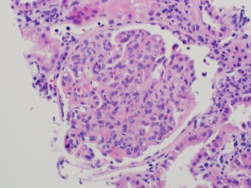
End

Previous Next
Multiple
  • Test Id: 198396adfe94861c89
  • QId: 167274
  • 66 of 96
  • Notes
  • Calculator
  • Feedback

A 35-year-old Caucasian male presents with persistent swelling of both legs associated with dark colored urine for two months. He went to an emergency room 2 months ago for these complaints and was told that he has some protein and blood in the urine. He was treated with levofloxacin for 3 days. There is no other past medical history. No history of skin rash or joint swelling. On examination,n the blood pressure was 130/85 mm H,g and there was bilateral 1+ pedal edema. The rest of the physical examination was normal. Urine analysis showed 3+ proteinuria, 10-15 RBCs per high-power field, and occasional RBC casts. BUN was 10 mg/dL, and serum creatinine was 0.9 mg/dL. Antistreptolysin was negative; C3 was decreased, and C4 was normal. Antinuclear antibodies, ANCA, hepatitis B and C serology were negative. 24-hour urine collection showed 2 g proteinuria, and a kidney biopsy was performed. Light microscopy showed increased mesangial matrix and cellularity, and the glomerular basement membrane appeared irregularly thickened. Silver stain revealed duplication of the glomerular basement membrane in multiple glomeruli. Immunofluorescence showed positive staining for C3, but negative for IgG, IgM and IgA. Electron microscopy revealed electron-dense deposits in the mesangium and subendothelial area. 

What is the most likely diagnosis?

A. Acute poststreptococcal glomerular nephritis
B. Diffuse proliferative lupus nephritis 
C. Dense deposit disease 
D. C3 glomerulopathy 
E.
Correct
The correct answer is D
C3 glomerulopathy

Explanation:

This patient has proteinuria, microscopic hematuria, hypertension and edema suggestive of acute nephritis of 2 months' duration. The classic prototype of acute nephritis is acute post-streptococcal glomerulonephritis. Typically, in acute post-infective glomerulonephritis, complement levels normalize within 6 weeks, and proteinuria and microhematuria generally resolve within about 6 weeks. Hence (Choice A) is wrong. This patient has hypocomplementemia and features of acute nephritis for 2 months,s suggesting that we should look for other causes of acute nephritis. 

Acute nephritis with low complement levels:
Post-infectious glomerulonephritis
Lupus nephritis
Membranoproliferativglomerulonephritisis
Mixed cryoglobulinemia
C3 Nephropathy

Acute nephritis with normal complement levels:
IgA nephropathy
Thin basement membrane disease
Hereditary nephritis
HSP
Mesangial proliferative glomerulonephritis
Lupus nephritis
Membranoproliferative glomerulonephritis
Mixed cryoglobulinemia
Dense deposit disease

This patient is a male, and ANA is negative. Lupus is mainly a disease affecting young women inthe third and fourth decades,e and most of them are ANA positive. A variety of glomerular pathologies are described, but immunofluorescence typically shows a 'full house' pattern with positive IgG, IgM, IgA and C3. ANA negativity and the absence of a full-house IF pattern make lupus nephritis (Choice B) unlikely.

Dense deposit disease is type II MPGN, and it mainly affects children and young adults. In most patients, there is a circulating C3 nephritic factor. Immunofluorescence microscopy demonstrates C3 deposits, and EM shows characteristic sausage-shaped, wavy deposits along the glomerular basement membranes (GBM) and mesangium. Lack of typical EM appearance makes dense deposit disease (Choice C) unlikely.

C3 Glomerulopathy is a morphologic variant of MPGN type I. In "C3 glomerulopathy", different light microscopy pathologies such as mesangioproliferative, membranoproliferative glomerulonephritis, diffuse proliferative glomerulonephritis, crescentic GN and sclerosing GN have been described—C3 usually low, C4 normal. In Sclerosing disease, C3 level can also be normal. The distinct feature is that IIF shows only C3 deposits, without any immunoglobulins. EM does not show sausage-shaped intramembranous deposits. Hence, the correct choice is (Choice D).

C3 glomerulopathy can present with varying degrees of proteinuria and azotemia and can progress to ESRD. It can recur after transplantation.

Educational Objective:
C3 glomerulopathy can present with varying degrees of proteinuria and azotemia and can progress to ESRD. It can recur after transplantation.
C3 glomerulopathy is a morphologic variant of MPGN type I. Its distinct feature is that IF shows only C3 deposits, without any immunoglobulins. EM does not show sausage-shaped intra-membranous deposits.

IMPORTANT BOARD EXAM ADVICE:

1. Differentiate between DDD and C3GN
2. Know the glomerulonephritis with low and normal complement levels.
3. BOARD QUESTIONS will be asked on DDD and C3GN, so know them well.

Copyright © ABIM Exam World
Created On: 09/12/2017
Last Modified: 10/13/2026

End

Previous Next
Multiple
  • Test Id: 198396adfe94861c89
  • QId: 167320
  • 67 of 96
  • Notes
  • Calculator
  • Feedback

A 25-year-old male comes to the physician complaining of flank pain and hematuria. He says the pain is constant and dull. There is no frequency, urgency or dysuria. He has a history of mental retardation and seizures. On physical exam, his blood pressure is 140/90 mm Hg, and his pulse is 80 bpm. He has multiple yellow papules on his nose and cheeks and numerous blanche, skin-colored spots on his face. A 2-3 cm hypopigmented macule is noted on the right arm. CT scan of the head was done as the patient presented with seizures. CT head was reported normal. CT scan of the abdomen shows bilateral hypodense fat-containing renal masses and cysts. 


What is the MOST likely diagnosis associated with these findings?

A.  Von Hippel Lindau
B.  Sturge Weber Syndrome
C. Tuberous Sclerosis
D.  Osler Weber Rendu
E.  Neurofibromatosis type 2
Correct
The correct answer is C
Tuberous Sclerosis

Explanation:
This patient’s skin lesions are consistent with sebaceous adenomas. The findings of mental retardation, sebaceous adenomas and seizures are most consistent with tuberous sclerosis. Tuberous sclerosis is associated with renal angiomyolipomas and renal cysts. Abdominal CT can diagnose these tumors because their fat density is lower than water. In patients with flank pain and hematuria, there is an increased likelihood of co-existing renal cysts. Based on the presentation and findings, this is tuberous sclerosis; the other choices are less likely, as explained below.

(Choice A) Bilateral renal cell carcinoma is associated with Von Hippel-Lindau disease. Imaging does not suggest renal cell carcinoma.
(Choice B) Leptomeningeal Angiomas are cerebral malformations commonly found in Sturge-Weber Syndrome. CT head is normal in this patient.
(C lO) Osler-Weber-Rendu is an autosomal dominant disease associated with angioectasias of the mucosal surfaces, as well as AVMs in the brain, GI tract and lung. Again, the clinical presentation and imaging do not suggest this diagnosis.
(Choice E) Neurofibromatosis type 2 is an autosomal dominant condition associated with acoustic neuromas, gliomas and ependymomas. The skin shows neurofibromas, but other associations favor tuberous sclerosis.

 ASSOCIATED FINDINGS IN TUBEROUS SCLEROSIS -- 
(This patient has the majority of these as bolded below)
 o      Bilateral renal angiomyolipomas (Fat-containing renal masses on CT scan)
 o      Renal Cysts
 o      Astrocytomas
 o      Cortical tubers
 o      Ash-leaf spots on skin
 o      Sebaceous adenomas on face
 o     Seizures
 o      Mental retardation

IMPORTANT TOPIC FROM RENAL BOARD POINT OF VIEW

Copyright © ABIM Exam World
Created On: 09/13/2017
Last Modified: 10/17/2026

End

Previous Next
Multiple
  • Test Id: 198396adfe94861c89
  • QId: 167322
  • 68 of 96
  • Notes
  • Calculator
  • Feedback

A 15-year-old boy is brought to the ER by his foster mother, who states that when she got home from work, she noticed he was acting very strange. He had slurred speech and seemed confused. He appeared very uncoordinated, and she was unsure whether he had fallen or hit his head. She states that he is somewhat a troubled boy but doesn’t know much about his history, as he has been in and out of the foster care system out of state. On physical exam, he is tachycardic and has tachypnoea. Pupils are dilated, but there is no nystagmus. Fundoscopy shows hyperemia of the optic disc is relatively uncooperative but not aggressive or hostile. When asked about suicidal thoughts, he responds only with inaudible mumbling. His foster mother left for work 10 hours prior and assumed he left for school. She is unsure when these symptoms began or what may have triggered them. P is 105/ min, BP is 140/90 mm Hg, RR is 28/min, and T is 97.1 F. Laboratory examination is as follows: 

Na   135 mEq/L                                            

K   5.0 mEq/L

CL   105 mEq/L

BUN  19 mg/dL

Cr   1.3 mg/dL         

HCO3  8 mEq/L  

Glucose  100 mg/dL         

pH   7.3          

pO2   90 mmHg

pCO2  22 mmHg

Measured serum osmolarity: 320 mmol/L

What is the next step in management?

A.  Gastric lavage
B.  N-acetylcystiene and activated charcoal
C. Fomepizole 
D. Fomepizole and Hemodialysis 
E.  Obtain serum levels of salycylate, methanol and ethylene glycol levels
Incorrect
The correct answer is D
Hemodialysis and fomepizole

Explanation: 
While this patient's case may appear to be vague, there are three strong indicators for the most likely diagnosis:
1)    This patient has metabolic acidosis with a profound Osmolar gap, which is a strong indicator of volatile alcohol related products. 
2)    Hyperemia of the optic disc (or other ocular symptoms), in addition to a profound anion gap, should make methanol toxicity a top differential diagnosis.  
3)    In addition to the serum anion gap, there is a serum Osmolar gap. (Measured Osm – Calculated Osm) where:

Calculated Osmolarity = 2(Na) + BUN/2.8 + Glu/18

***Anytime there is an osmolar gap in metabolic acidosis, your differentials can be narrowed to volatile alcohol-related substances. ***  --------------->    IMPORTANT ABIM BOARD POINT

The best treatment for methanol and ethylene glycol is to remove the toxic byproducts from circulation. The definitive therapy is Hemodialysis. Fomepizole is given to block alcohol dehydrogenase, thereby stopping the formation of toxic byproducts. However, it does not remove them from circulation; only dialysis can.

(Choice A) Gastric lavage is rarely the correct answer, especially with ingestion of an unknown substance. Caustic substances can cause further damage during gastric lavage. Esophageal perforation is potentially a fatal risk.

(Choice B) N-acetylcysteine is the treatment for acetaminophen toxicity; however, timing is important, and drug levels should be drawn first to determine the need for treatment. If ingestion occurred more than 24 hours earlier, treatment has no effect and is therefore not given.

(Choice C, D) Refer to the explanation section above.

(Choice E) When suspicion for methanol poisoning is high, treatment should not wait for diagnosis confirmation. Ocular involvement is an ominous sign, and the risk of blindness increases. In this case, the benefits outweigh the risk of treating before confirming the diagnosis.


Copyright © ABIM Exam World
Created On: 09/13/2017
Last Modified: 10/17/2026

End

Previous Next
Multiple
  • Test Id: 198396adfe94861c89
  • QId: 167339
  • 69 of 96
  • Notes
  • Calculator
  • Feedback

All of the following help predict AV Graft stenosis EXCEPT:

A. Hyperpulsatility of the AV Graft
B. Ruduced pulse augmentation
C. Increased bleeding and clots 
D. Decreased KT/V
E. Decreased Blood Flow rate
F. Surveillance of the graft
Incorrect
The correct answer is F.

Surveillance of the graft

Explanation:

Intra-graft or venous outflow stenosis – A strong pulse in the AV graft (hyperpulsatility) suggests increased resistance, as occurs with venous outflow stenosis. Pulse intensity is directly proportional to stenosis severity. For this reason, in an AV graft, hyperpulsatility can indicate impending AV graft stenosis.

Arterial stenosis – REDUCED PULSE AUGMENTATION suggests inflow stenosis due to stenosis of the arterial anastomosis or of the feeding artery. Although pulse augmentation is commonly performed, it is less sensitive for detecting inflow stenosis in AV grafts than in AV fistulas.

INCREASED BLOOD CLOTS, DECREASED KT/V, and DECREASED BLOOD FLOW RATE are all suggestive of impending AV graft stenosis. Patients often have dilated collateral veins over the arm and chest wall (very important to examine your hemodialysis patients after they take off their shirts), suggesting central venous stenosis. The classic physical finding in a patient with significant central venous stenosis is diffuse upper extremity edema. Subcutaneous collateral veins are frequently evident over the chest. Swelling and collateral veins are caused by generalized venous hypertension of the extremity, which occurs in central lesions but is rare with peripheral lesions. 

SURVEILLANCE  does not predict AV Graft stenosis. It is not a sensitive or specific modality for accurate detection.

Copyright © ABIM Exam World
Created On: 09/14/2017
Last Modified: 10/17/2026

End

Previous Next
Multiple
  • Test Id: 198396adfe94861c89
  • QId: 167340
  • 70 of 96
  • Notes
  • Calculator
  • Feedback

A 40-year-old pleasant African man with ESRD secondary to FSGS started automated peritoneal dialysis. His prescription includes 2.5 L and 3 exchanges over 8 hours at night, with a last fill of 2 L. His urine output is 1000 mL/day. Typical ultrafiltration on the cycler is 1000 mL. The average drain volume during the day dwell was 1500 mL before going on the cycler at night. 

He came with complaints of lower abdominal wall edema extending to the scrotum over the past 5 days. Without any change in the dialysis prescription, his drain volume before going on the cycler dropped to 900 mL, and the ultrafiltration volume on the cycler came down to 100 mL. He reports no pain with fill or drain. 

What is the next step?


A. Chest X ray PA and lateral view 
B. Drain the fluid middle of the day to reduce the dwell time
C. PD catheter manipulation
D. Abdominal CT scan with contrast in the dialysate
E. Switch to hemodialysis 
F. Pleurodesis 
Correct
The correct answer is D. 

Abdominal CT scan with contrast in the dialysate.e

Explanation:

Typical ultrafiltration failure from peritoneal membrane failure presents gradually. More often, we see apparent ultrafiltration failure for other reasons. 

Catheter malfunction can present with problems with inflow, outflow or both. A kink in the catheter can cause problems with inflow and outflow. 

Omentum or blood clots clogging the catheter can cause outflow problems only. Fibrin clots, constipation with a loaded rectum, and a displaced PD catheter also cause outflow obstruction. A KUB X-ray is very helpful in diagnosing a displaced catheter.   

Fluid leak into the abdominal wall causes swelling in the lower abdomen extending down to the scrotum or mons pubis and a decrease in drain volume. Usually, this can be treated by temporarily doing low-volume PD or transferring to HD for about 6 weeks. Leaks starting a few weeks after initiating PD usually do not respond to this approach and often require placement of a new PD catheter.

A patent tunica vaginalis testis will cause UNILATERAL OR BILATERAL SCROTAL SWELLING without much swelling of the abdominal wall, sometimes associated with a decrease in the drain volume. A CT scan of the abdomen with ‘IV’ contrast into the peritoneal fluid can help in diagnosing the leak into the abdominal wall and scrotal leak. Patent tunica vaginalis testis requires surgery. Generally, hemodialysis is not required after the surgery, and low-volume peritoneal dialysis can be resumed. This patient presents with signs of leak of fluid into the abdominal wall. A chest X-ray is not required, so (Choice A) is wrong.

Draining during the day helps if the membrane is very permeable, with a very high D/P creatinine ratio on PET. This does not help with an abdominal wall leak of dialysate. Therefore, (Choice B) is also wrong.

PD catheter manipulation (Choice C) is wrong because the patient does not have a displaced PD catheter based on the information given.

(Choice D) is the correct answer. If the clinical presentation is very convincing, most patients are treated by reducing the dialysate volume and, if possible, keeping the abdomen dry during the day. Sometimes these patients need to be switched to hemodialysis for 4-6 weeks before PD is resumed. Hence, a temporary switch to HD would be a correct answer, but an unqualified switch to HD, as stated in answer E, would be wrong. Therefore, (Choice E) is the wrong or less appropriate answer.

ABDOMINAL ADHESIONS cause a decrease in the drain volume as well and require surgical treatment with low-volume PD or temporary HD after that.
A 
DIAPHRAGMATIC LEAK into the pleural space presents with cough and shortness of breath without peripheral signs of fluid overload, a few weeks after starting peritoneal dialysis (not the presentation in this patient). Usually, such patients present with right-sided pleural effusion clinically and on chest X-ray (PA and lateral view. These patients can be treated with temporary transfer to HD, pleurodesis and return to PD 4-6 weeks later. Permanent switch to hemodialysis without pleurodesis is another option.


TREATMENT OPTIONS OF VARIOUS ABDOMINAL/CHEST PROBLEMS AS MENTIONED ABOVE IN PATIENTS ON PERITONEAL DIALYSIS ARE FREQUENTLY TESTED ON BOARDS. PLEASE KNOW THE TREATMENT OPTIONS.


Copyright © ABIM Exam World
Created On: 09/14/2017
Last Modified: 10/17/2026

End

Previous Next
Multiple
  • Test Id: 198396adfe94861c89
  • QId: 167345
  • 71 of 96
  • Notes
  • Calculator
  • Feedback

Cyclosporine nephrotoxicity in a renal transplant recipient is associated with all of the following renal biopsy findings EXCEPT:

(THIS PICTURE BELOW IN LOW POWER SHOWS ONE OF THE CLASSICAL FINDINGS IN CSA TOXICITY)


A. Interstitial Fibrosis  
B. Tubular atrophy 
C. Endothelial cell swelling 
D. Glomerular thrombin deposits
E. Glomerular basement membrane thickening 
F. Double contours of the GB 
Incorrect
The correct answer is D.

Glomerular thrombin deposits

Explanation:

In a patient with suspected cyclosporine nephrotoxicity, renal biopsy reveals obliterative arteriolopathy (classically in the afferent renal arterioles), suggesting primary endothelial damage and subsequent endothelial cell swelling, which may persist for months with elevated cyclosporine blood levels. This is also associated with a thickened glomerular basement membrane and a double-contour pattern. In fact, according to BANF, a thickened glomerular basement membrane and double-contour pattern are most suggestive of chronic allograft nephropathy (CAN), also called transplant glomerulopathy. 

The other renal biopsy findings of cyclosporine nephrotoxicity include ischemic collapse or scarring of the glomeruli, vacuolization of the tubules, FSGS, and focal areas of tubular atrophy and interstitial fibrosis (producing a picture of"“ZEBR"” or""STRIPE"" fibrosis). These changes are seen with both low-dose and higher-dose cyclosporine therapy. However, they seem to correlate earlier with higher doses. 

(THE ABOVE PICTURE IN THE UPPER HALF SHOWS TUBULAR ATROPHY APPEARING DARK AND REDDISH ALTERNATING WITH LIGHT BLUE AREAS OF INTERSTITIAL FIBROSIS GIVING A "STRIPE" OR" " ZEBRA APPEARANCE)

THE PICTURE BELOW SHOWS TUBULAR ATROPHY, VACUOLIZATION OF THE TUBULES AND ISCHEMIC CHANGES:


Mild arteriolar hyalinosis at six months appears to be associated with high doses of cyclosporine and was reversible. However, after more than a year, irreversible severe arteriolar hyalinosis and glomerulosclerosis were observed, despite decreased doses and trough levels of cyclosporine.

Glomerular thrombin deposits are typically seen in patients with Lupus, antiphospholipid syndromes and other vasculitides. It is typically not seen in cyclosporine nephrotoxicity.

Copyright © ABIM Exam World
Created On: 09/14/2017
Last Modified: 10/17/2026

End

Previous Next
Multiple
  • Test Id: 198396adfe94861c89
  • QId: 167347
  • 72 of 96
  • Notes
  • Calculator
  • Feedback

A 68-year-old African American male with a known history of diabetes mellitus,  hyperlipidemia, CVD, and GERD presents to the ER with chest pain and shortness of breath. The patient says the pain is crushing and radiates to his left arm. It woke him from sleep 30 minutes ago. He took his antacids and 4 tablets of nitroglycerin, and the pain got better. His current medications include low-dose aspirin, metoprolol, glyburide, pioglitazone, lisinopril, simvastatin, and omeprazole. He recently suffered an upper respiratory infection for which he was given Levofloxacin. Vitals: BP 160/95 mm Hg, T 98.1, HR 110. He has no leg edema and no jugular venous distention. EKG shows LVH by voltage criteria with an isoelectric ST segment. Serial troponin levels are normal. A stress test showed evidence of stress-induced ischemia in the anterior leads. He is scheduled for cardiac catheterization the next day. Laboratory results are as follows:

Serum Chemistry:

Na   143 mEq/L

K   3.5 mEq/L

Cl   101 mEq/L

HCO3  22 mEq/L

BUN   35 mg/dL

Cr   3.0 mg/dL

Glucose  110 mg/dL 

What would you do to PROTECT the kidneys before doing the cardiac catheterization?

A. Dopamine IV infusion before and after the  procedure.
B. Nifedipine.
C. Isotonic sodium bicarbonate infusion before and after the procedure.
D. N Acetyl Cysteine before and after the procedure
E. Start mannitol IV before the procedure
F. Left ventriculogram quickly with coronary angiogram
Correct
The correct answer is C.

Isotonic sodium bicarbonate infusion before and after the procedure. N-acetylcysteine is also used, but its efficacy in studies is unproven.

Explanation:

Contrast nephropathy usually presents as a rise in creatinine 24-48 hrs after IV radiocontrast exposure.

Other features of CONTRAST NEPHROPATHY are:
  • Low fractional excretion of sodium (FeNa) below 1%.
  • Iodinated contrast agents can cause false-positive proteinuria so that proteinuria may be overestimated.
  • It is reversible most of the time.
  • UA may show muddy brown granular casts, tubular epithelial cell casts and numerous tubular epithelial casts.

Contrast-associated nephropathy has to be differentiated from atheroembolic disease, which presents with deterioration in kidney function days to weeks after the procedure,  embolic lesions (such as digital ischemia of the toes) or livedo reticularis, transient eosinophilia and hypocomplementemia,  protracted course with frequently little or no recovery of renal function, and kidney biopsy showing the typical biconvex, needle-shaped clef"s ("gh" sts") within the occluded vessel from the space occupied by the cholesterol crystal that dissolves during the processing of the tissue, givin' a ‘fish m'uth’ appearance.

(Choice A) There is no evidence to support that dopamine infusion is beneficial. In fact, it may be harmful.

(Choice B) No evidence supports that nifedipine is beneficial.

(Choice C) Studies suggest that infusion of isotonic saline or half-normal saline may be beneficial. Since alkalization may protect against free radical injury, sodium bicarbonate may be superior to isotonic saline. In patients who tolerate extra volume (i.e. those who are not volume overloaded or those who are not in heart failure), it would be reasonable to infuse isotonic sodium bicarbonate or saline.

(Choice D) Some studies suggest that oral N-acetylcysteine before or after the procedure reduces the risk of contrast nephropathy. Other studies suggest it is not beneficial. N-acetyl cysteine is believed to work through its vasodilator and antioxidant effects. Because N-acetylcysteine is safe and inexpensive, many physicians would administer it prophylactically as well, even though results are conflicting and expensive.

(Choice E) There are studies to suggest that prophylactic conflictosemide is harmful.
    
(Choice F) Left ventriculogram should be avoided in patients at high risk of developing contrast nephropathy to minimize the quantity of IV contrast needed.

RISK FACTORS for contrast nephropathy are :
  • Chronic kidney disease
  • Diabetes mellitus
  • Congestive heart failure
  • Myeloma kidney
  • Hypotension

Educational Objective:

1. Identify and diagnose contrast nephropathy

2. Prevention of contrast nephropathy

Copyright © ABIM Exam World
Created On: 09/14/2017
Last Modified: 10/17/2026

End

Previous Next
Multiple
  • Test Id: 198396adfe94861c89
  • QId: 16521
  • 73 of 96
  • Notes
  • Calculator
  • Feedback

As per the JNC VIII committee recommendation, for individuals who are part of the African American population, including those with diabetes, the initial treatment should include a thiazide-type diuretic or calcium channel blocker (CCB).  

Which of the following thiazide-type diuretics is preferred as the initial antihypertensive therapy?


A. Hydrochlorthiazide 
B. Chlorthalidone 
C. Indapamide 
D. Metalazone 
E. Polythiazide 
Correct
The correct answer is B 
Chlothalidone

Explanation:

• Chlorthalidone at the same dose is approximately 1.5 to 2 times as potent as hydrochlorothiazide.

• Chlorthalidone has a longer duration of action than hydrochlorothiazide.

• There are no randomized clinical trials that directly compare outcomes in hypertensive patients treated with hydrochlorothiazide versus chlorthalidone. A multiple treatment (network) meta-analysis of nine trials including 50,946 patients was conducted in which hydrochlorothiazide and chlorthalidone were indirectly compared by evaluating their efficacy against common comparative drugs (ACE inhibitors were compared with hydrochlorothiazide in the ANBP2 trial and with chlorthalidone in the ALLHAT trial). The major findings of this meta-analysis showed that chlorthalidone significantly reduced the risk of cardiovascular events compared to hydrochlorothiazide (relative risk 0.79, 95% CI 0.72 to 0.88) and heart failure (relative risk 0.77, 95% CI 0.61 to 0.98). The authors calculated that 27 patients would need to be treated with chlorthalidone instead of hydrochlorothiazide over five years to prevent one cardiovascular event. Chlorthalidone remained superior to hydrochlorothiazide even after the meta-analysis was controlled for achieved office systolic blood pressure (relative risk for cardiovascular events 0.82, 95% CI 0.70 to 0.97). This finding may reflect chlorthalidone's longer duration of action and lower nocturnal blood pressure.

• Multiple Risk Factor Intervention Trial (MRFIT) - Men Hypertensive 2392 were treated with chlorthalidone, and 4049 were treated with hydrochlorothiazide. During six years of follow-up, cardiovascular events (defined as myocardial infarction, stroke, coronary artery bypass surgery, heart failure, left ventricular hypertrophy, peripheral artery disease, or angina) were significantly less common with chlorthalidone compared with hydrochlorothiazide (hazard ratio 0.79, 95% CI 0.68 to 0.92). Through the course of the study, systolic blood pressure and LDL cholesterol levels were also lower with chlorthalidone compared with hydrochlorothiazide. 

• Indapamide is a thiazide-like diuretic and has a half-life of 14-16 hours. This drug has been used in the HYVET trial alone or in combination with perindopril in the treatment of hypertension in patients more than 80 years old. The study showed a 30% reduction in stroke, a 39% reduction in the rate of death, a 21% reduction in death from any cause, a 23% reduction in CV death and a 64% reduction in the rate of heart failure. The trial showed that careful BP lowering in the very elderly is beneficial. Indapamide is not preferred over chlorthalidone.

• Metolazone - There are many studies with metolazone available. An important additional property is its effectiveness as a diuretic at lower GFR values. The duration of action is about 24 hours.
It is not preferred over chlorthalidone.

Copyright © ABIM Exam World
Created On: 09/20/2017
Last Modified: 10/17/2026

End

Previous Next
Multiple
  • Test Id: 198396adfe94861c89
  • QId: 16524
  • 74 of 96
  • Notes
  • Calculator
  • Feedback

Which of the following antihypertensive medications resulted in better blood pressure control and cardiovascular outcomes when combined with an ACE inhibitor?


A. Beta blocker 
B. Calcium channel blocker
C. Angiotensin Receptor Blocker 
D. Diuretics 
Correct
The correct answer is B
Calcium channel blocker 

Explanation:

The ACCOMPLISH trial has clearly shown that an ACE inhibitor combined with a Calcium channel blocker (amlodipine) is better than an ACE inhibitor combined with a diuretic (hydrochlorothiazide). This study did not involve beta blockers or angiotensin receptor blockers. 

The ACCOMPLISH trial was the first major clinical trial addressing the issue of combination therapy in 11,000 patients who were at high cardiovascular risk (with prior CAD, diabetes, and/or CKD). The patients were randomly assigned to combination therapy with Benazepril (20 mg/day) combined with either amlodipine (5 mg/day) or hydrochlorothiazide (5 mg/day).

The Benazepril-Amlodipine group had significantly lower mean blood pressure, and the primary endpoint was also significant in this group. There was a similar benefit with the treatment combination of benazepril-amlodipine in the secondary endpoint of cardiovascular death compared to the group treated with a combination of amlodipine and hydrochlorothiazide

Copyright © ABIM Exam World
Created On: 09/20/2017
Last Modified: 10/17/2026

End

Previous Next
Multiple
  • Test Id: 198396adfe94861c89
  • QId: 165212
  • 75 of 96
  • Notes
  • Calculator
  • Feedback

The ACCOMPLISH trial is the first major trial addressing the issue of combination therapy in 11,506 patients who were at high cardiovascular risk. The goal blood pressure was <130/80 mm Hg in patients with diabetes or impaired renal function, and <140/90 mm Hg in patients with prior cardiovascular disease.

Which of the following combinations of blood pressure medications was the best in reducing cardiovascular events and slowing the progression of nephropathy in patients with hypertension who were at high risk for such events?

A. ACEI + Diuretics
B. ACEI + CCB 
C. ACEI + Beta-blocker 
D. CCB + Beta-blocker 
Correct
The correct answer is B
ACEI + CCB

Explanation:

• The Avoiding Cardiovascular Events through Combination Therapy in Patients Living with Systolic Hypertension (ACCOMPLISH) trial showed that initial antihypertensive therapy with benazepril plus amlodipine was superior to benazepril plus hydrochlorothiazide in reducing cardiovascular morbidity and mortality.

• ACCOMPLISH ACCOMPLISH was a double-blind, randomized trial undertaken in five countries (USA, Sweden, Norway, Denmark, and Finland). 11 506 patients with hypertension who were at high risk for cardiovascular events were randomly assigned via a central, telephone-based interactive voice response system in a 1:1 ratio to receive benazepril (20 mg) plus amlodipine (5 mg; n=5744) or benazepril (20 mg) plus hydrochlorothiazide (12.5 mg; n=5762), orally once daily. Drug doses were force-titrated for patients to attain recommended blood pressure goals.

• The primary endpoint was the composite of death from cardiovascular causes, nonfatal myocardial infarction, nonfatal stroke, hospitalization for angina, resuscitation after sudden cardiac arrest, and coronary revascularization.

• The benazepril-amlodipine combination was superior to the benazepril-hydrochlorothiazide combination in reducing cardiovascular events in patients with hypertension who were at high risk for such events.

Copyright © ABIM Exam World
Created On: 09/20/2017
Last Modified: 10/17/2026

End

Previous Next
Multiple
  • Test Id: 198396adfe94861c89
  • QId: 165215
  • 76 of 96
  • Notes
  • Calculator
  • Feedback

An 18-year-old male is brought to the renal clinic for evaluation of hypertension. There is no history of edema of the feet, puffiness of the face, hematuria, oliguria or recurrent urinary tract infections. He is an active football player, but lately he complains of weakness and muscle cramps. His blood pressure for the last 3-4 years has always been borderline high and during this visit was as noted below.

Physical examination shows: Pulse 100/min; all peripheral pulses are well felt. BP 150/100 mm Hg, Temp. 37.4 degrees C. He is pale. His growth is stunted. His cognitive function is normal, and no focal neurological deficits are noted. Other systems are unremarkable. Laboratory examination is as follows:

Hemoglobin   13.5 g/dL

Hematocrit   42%

Platelet Count   150,000 mm3

White Blood Cells  8,000 mm3


Urinalysis: 

Protein     100 mg/24 h

Glucose       None

Red Blood Cells      None

White Blood Cells     None

Leukocyte Esterase   Negative

Nitrites     Negative


Chemistry: 

BUN   13.5 mg/dL

Creatinine  0.9 mg/dL

Sodium   140 mEq/L

Potassium  5.8 mEq/L

Bicarbonate  18.5 mEq/L

Chloride   112 mEq/l

Calcium   9.2 mEq/L

Phosphorus  3.2 mg/dL

Aldosterone  5 ng/mL

PRA   <1.0 ng/dL/hr


Glucose   100 mg/dL

HbA1C    5.30%

S. Osmolality   282 mOsmol

Uric Acid   5.3 mg/dL

ESR    9.8 mm/h


ABG: 

pH   7.25

PCO2  32 mm Hg

HCO3 16 mEq/L

PO2   90 mm Hg

U: NNormal-sizedkidneys, no hydronephrosis 

Considering the history and the laboratory findings, which of the following is most likely the cause of hypertension in this patient?


A. Liddle's Syndrome 
B. Gordon's Syndrome 
C. Congenital adrenal hyperplasia 
D. Glucocorticoid-remediable aldosteronism 
E. Mineralocorticoid receptor activating mutation 
Correct
The correct answer is B
Gordon’s syndrome

Explanation:

Gordon’s syndrome - A young male with hypertension, hyperkalemia, hyperchloremic metabolic acidosis, normal renal function, low renin, and low aldosterone is highly characteristic of Gordon’s syndrome. This syndrome is also known as pseudohypoaldosteronism type II. Recently, it has been reported that Gordon’s syndrome is linked to chromosomes 1, 12, and 17. Hyperkalemia and hypertension in Gordon’s syndrome are caused by abnormalities in WNK kinases, proteins that are localized to the distal nephron and affect the thiazide-sensitive Na-Cl Cotransporter. Mutations in WNK4 or WNK1 increase chloride reabsorption in the distal nephron, reducing lumen electronegativity and lowering the driving force for potassium secretion. These mutations also decrease expression of the potassium channels through which potassium enters the collecting duct. Thus, mutant WNK4 increases sodium chloride reabsorption in the distal tubule and decreases potassium secretion in the collecting duct, resulting in sodium retention, decreased potassium excretion, hypertension, hyperkalemia, and metabolic acidosis. Volume expansion suppresses renin and aldosterone. 

(Choice A) Liddle’s syndrome presents as hypertension in childhood. It is usually resistant to standard antihypertensive drugs. This is due to a gain-of-function mutation on chromosome 16 affecting the beta or gamma subunit of the ENaC channel in the collecting duct of the nephron. This results in sodium retention and volume expansion, causing hypertension and hypokalemia. Volume expansion suppresses renin and aldosterone. These patients will have hypertension, hypokalemia, metabolic alkalosis, low renin and low aldosterone. 

(Choice C) Congenital adrenal hyperplasia presents in childhood with hypertension and virilization in girls and precocious puberty in boys. The other features include primary amenorrhea, delayed sexual maturation, ambiguous genitalia, with clitoral enlargement in girls.Deficiencies in  11-beta-hydroxylase (CYP11B1) and 17-alpha-hydroxylase (CYP17) lead to excess deoxycorticosterone, whichcauseso hypertension through mineralocorticoid action. Biochemically, it is characterized by metabolic alkalosis, low renin and low aldosterone.

(Choice D) Glucocorticoid-remediable aldosteronism (GRA) is also known as familial hyperaldosteronism type I. In this disorder, a gene mutation affects the 11-beta-hydroxylase gene (CYP11B2) and the aldosterone synthase gene (CYP11B2). Patients with GRA have ectopic aldosterone synthesis occurring in the zona fasciculata under the direct influence of ACTH rather than renin. GRA presents with hypertension, hypokalemia, metabolic alkalosis, low renin and high aldosterone. GRA resembles a primary hyperaldosteronism state with excessive urinary potassium loss. Look for a clue of family history of CVA or brain haemorrhage in the stem of the question.

(Choice E) A mineralocorticoid receptor-activating mutation is a very rare autosomal dominant disorder. The mutation lies in the mineralocorticoid receptor, causing it to be activated by steroids lacking 21–hydroxyl groups, such as progesterone and spironolactone. Both normally antagonize the receptor but have opposite effects on this mutant receptor. It usually presents as early-onset hypertension in females. It may get accelerated during pregnancy due to elevated levels of progesterone. Biochemically,  it is characterized by hypokalemia, metabolic alkalosisis low renin and low aldosterone.

Educational objective:

IF YOU KNOW THESE 5 POINTS FOR THESE 5-6 SYNDROMES,THENS YOU CAN ANSWER ALL NEPHROLOGY BOARD QUESTIONS ASKED ON THESE SYNDROMES:

Blood pressure - Normotension or hypertension
Potassium        - Hyperkalemia or hypokalemia
Acid-Base         - Acidosis or alkalosis
Renin                - Low or High
Aldosterone      - Low or High

Copyright © ABIM Exam World
Created On: 09/20/2017
Last Modified: 10/17/2026

End

Previous Next
Multiple
  • Test Id: 198396adfe94861c89
  • QId: 165230
  • 77 of 96
  • Notes
  • Calculator
  • Feedback

A 56-year-old male was brought to the emergency room with drowsiness and lethargy. He has had these symptoms for the past 2 days. He complains of a recent history of anorexia, nausea, and vomiting. He has diabetes mellitus and has been on glimepiride 1 mg daily for the last 4 years. One week ago, he had decreased vision with redness in his right eye. He was treated by his ophthalmologist with drops,s which seem to have resolved the problem. He currently takes cholecalciferol weekly for osteoporosis. On physical examination, his pulse is 80/min, blood pressure is 140/90 mm Hg, respiratory rate is 20/min, and temperature is 97.7°F. The patient appears drowsy but shows no focal neurological deficits. Review of systems is otherwise unremarkable. Urinalysis is positive for glucose and negative for proteinuria, WBCs and RBC casts. A24-hour urinary protein collection shows proteinuria of 3.5 g/day. Further labs reveal:

Hemoglobin  8 gm%

Hct    24%

MCV   85

WBC   7800/ml

PMN   80%

Lymphocytes  20%

ESR   80 mm/hr

Na    145 mEq/L

BUN   80 mg/dL

Cr    1.8 mg/dL

CL    115 mEq/L

HCO3   25 mEq/L

Uric acid   5.8 mg/dL

Ca    14 mg/dl

PO4    2.8 mg/dL

Total Protein  7.8 gm/dL

Albumin   3.5 mg/dL.

Vitamin D  40 ng/ml

PTH   10 pg/ml

Which of the following is most likely the cause of his hypercalcemia?


A.  Vitamin D toxicity
B. Primary hyperparathyroidism
C.  Multiple myeloma.
D. Sarcoidosis
Correct
The correct answer is C.

Multiple myeloma

Explanation:

Multiple myeloma results from the proliferation of plasma cells producing a monoclonal immunoglobulin.

The diagnosis of multiple myeloma is supported by the presence of many of the following factors:
  • Advanced Age
  • Backache and Bone pain
  • Anemia
  • High ESR
  • Urine protein negative by dipstick and positive by other tests is characteristic of light chain proteinuria.
  • High Total protein
  • Low anion gap
  • Hypercalcemia and hyperuricemia favor the diagnosis of multiple myeloma.

(Choice A) This patient has received Vitamin D treatment. Vitamin D toxicity can give rise to hypercalcemia. Serum vitamin D3 levels will be high. PTH will be low. Patient’s vitamin D level of 40ng/ml (normal range is 30-100ng/ml) does not favor Vitamin D toxicity.

(Choice B) Primary hyperparathyroidism is the commonest cause of hypercalcemia among outpatients. It is characterized by elevated PTH. The patient’s PTH level of 10 pg/ml rules out primary hyperparathyroidism.

(Choice D) Sarcoidosis is a multisystem granulomatous disorder of unknown cause. Hypercalcemia and hypercalciuria in sarcoidosis are associated with high vitamin D levels. This patient has low vitamin D levels, so it is unlikely to be the cause. There is also no history or findings to suggest sarcoidosis.

Copyright © ABIM Exam World
Created On: 09/20/2017
Last Modified: 10/17/2026

End

Previous Next
Multiple
  • Test Id: 198396adfe94861c89
  • QId: 165233
  • 78 of 96
  • Notes
  • Calculator
  • Feedback

A 25-year-old female is referred by her primary care provider for evaluation of hypertension and hypokalemia. Her primary care provider has already started her on oral potassium despite a potassium level of 2.8 mEq/L. Despite amlodipine and lisinopril, her blood pressure is 154/96 mm Hg. There is no renal bruit. Systemic and fundus examinations are normal. Her mother was also diagnosed with hypertension at an early age. Her brother died of a cerebrovascular accident 2 years ago. Laboratory findings are as follows:

Na   140 

Potassium 2.8 

Chloride   100 

HCO3    26

BUN   15 

Creatinine  0.8 

Glucose    110

TSH and Cortisol are normal

ACTH    elevated

Renin   0.7 (Low)

Aldosterone  48 (elevated)


Urinalysis:

Sodium   240 mEq/D

Potassium  98 mEq/D

Urinary 18-OH Cortisol and 18-oxocortisol are elevated.

The most appropriate treatment for this patient is:

A. Steroids
B. Spironolactone 
C. Steroids + Spironolactone
D. Amiloride 
Correct
The correct answer is C.

Steroids + Spironolactone

Explanation:

This patient has (GRA, glucocorticoid-responsive aldosteronism). These are typically young patients presenting with hypertension and hypokalemia. There is a strong family history of early-onset hypertension along with a history of fatal CVA or hemorrhagic strokes. These patients typically have low Renin and high aldosterone, mimicking a state of primary hyperaldosteronism. As a result, they have high urinary potassium losses, as seen in this patient. (Urinary potassium of more than 40mEq/D is considered increased losses). Increased ACTH and 18-OH cortisol further support the diagnosis of GRA.

Steroids alone are used in the treatment of pregnancy-associated mineralocorticoid excess and congenital adrenal hyperplasia. Steroids alone in this patient will suppress ACTH, but this patient also needs an aldosterone antagonist for the hyperaldosteronism and to reduce urinary potassium losses. Hence, the right answer is steroids plus spironolactone.

Spironolactone alone is not sufficient, as steroids are needed to suppress the increased ACTH state. 

Amiloride blocks the ENaC channel in Liddle syndrome.

PLEASE NOTE THAT "G" AND "S " ARE FREQUENTLY ASKED QUESTIONS IN NEPHROLOGY BOARD EXAMS

PLEASE REVIEW GLUCOCORTICOID-REMEDIABLE ALDOSTERONISM WELL. 

Copyright © ABIM Exam World
Created On: 09/20/2017
Last Modified: 10/17/2026

End

Previous Next
Multiple
  • Test Id: 198396adfe94861c89
  • QId: 165242
  • 79 of 96
  • Notes
  • Calculator
  • Feedback

A 60-year-old with recently diagnosed colon cancer and diabetes presents with bilateral pedal edema; BP is 120/80 mm Hg. Urinalysis showed  4+ protein, no RBCs or WBCs, and 8-10 Hyaline casts. His BUN is 20, Creatinine is 1 mg/dL, and albumin is 2 grams/dL. A 24-hour urine collection showed 10 grams of protein. The patient undergoes a kidney biopsy. The EM is shown below :


What is the most likely diagnosis?

A.  Crescentic GN
B.  IgA nephropathy
C.  Minimal change disease
D.  Focal segmental glomeruloscerosis
E.  Membranous nephropathy
F. Diabetic nephropathy
Correct

The correct answer is E

Membranous Nephropathy.

Electron microscopy shows subepithelial electron-dense deposits, as classically seen in membranous nephropathy. If in the question there is a suggestion of colon, breast, or lung cancer, then the glomerulopathy is usually membranous. Then look for other histopathological findings that confirm the diagnosis: subepithelial electron-dense deposits.

Explanation:

BOARD POINT - FAMILIARIZE YOURSELF WITH THESE ASSOCIATIONS :

1. Solid cancers (colon, breast, lung, renal) plus proteinuria = Membranous nephropathy

2. Hodgkin's lymphoma plus proteinuria = Minimal change disease

3. HIV plus proteinuria = Focal segment glomerulosclerosis FSGS

4. Pamidronate plus protenuria = FSGS (rare)

5. Myeloma, no albuminuria on dipstick, but proteinuria on protein/creatinine ratio or 24-hour urine: Light chain nephropathy

6. Myeloma with nonspecific proteinuria (on dipstick, urine protein/creatinine ratio and 24-hour urine): Light chain nephropathy or amyloidosis.


BOARD POINT - FAMILIARIZE YOURSELF WITH THESE HISTOPATHOLOGY ASSOCIATIONS FOR VARIOUS GLOMERULAR DISEASES


Copyright © ABIM Exam World
Created On: 09/20/2017
Last Modified: 10/17/2026

End

Previous Next
Multiple
  • Test Id: 198396adfe94861c89
  • QId: 165243
  • 80 of 96
  • Notes
  • Calculator
  • Feedback

You are the nephrologist on call. The  ER calls you for an 18-year-old female who is complaining of vomiting and diarrhea. Her serum sodium is 116 mEq/L and Serum potassium is 5.9 mEq/L. On physical examination, the patient is drowsy; pulse is 126/min, BP is 90/60 mm Hg, and RR is 32/min. Her chest is clear. Her heart sounds are normal, with no murmur. The patient is drowsy but arousable, with no focal neurological deficit. Laboratory findings are as follows:

Hb    16 gm/dl 

WBC   12,800/cmm 

Polymorph  46% 

Lymphocytes  16% 

Eosinophils  4%

Monocytes  4%

Platelets   2,40,000/cmm.

CL    70 mEq/L

BUN  10 mg/dl

Creatinine  0.5 mg/dl

Na    116 mEq/L

K    5.8 mEq/L


Urinalysis:

pH    6.4

Protein  trace

Glucose  absent

Microscopic: occasional WBCs & RBCs

Urinary Na  90 mEq/L

Urinary K         20 mEq/L

ABG    

PH                   7.32

PCO2   36 

HCO3   20 mEq/L

PaO2   92

O2 saturation  98%

S. Cortisol  6.00 mg/dl

TSH   3.5 IU/m (Normal 0-5 IU/m ).

Both plasma Renin and Aldosterone are high. 

Which of the following conditions is most likely with these findings?


A. Type 4 RTA 
B. Pseudo-hypo-aldosteronism Type 1 
C. Gordon's syndrome
D.  Diarrhea
Incorrect
The correct answer is B.
 Pseudo-hypoaldosteronism type 1

Explanation:

This patient has hyponatremia with high urinary sodium, high serum potassium, low urinary potassium, and normal adrenal function. The serum aldosterone level is high, suggesting target-organ resistance to aldosterone. These findings are characteristic of pseudohypoaldosteronism type 1 (PHA type 1)

PHAtype 11 is a rare hereditary disorder, characterized by generalized resistance to aldosterone action. It presents with 
1. Salt wasting 
2. Hypovolemia 
3. Normotension
4. Metabolic acidosis 
5. Hyperkalemia
6. High Renin and Aldosterone.  

NEPHROLOGY BOARD EXAM TAKERS REMEMBER THESE 6 POINTS FOR PHA TYPE 1

These are two different modes of inheritance:
1] Autosomal recessive.
2] Autosomal dominant.

Autosomal recessive affects the epithelial sodium channel and other target organs, such as the kidney, colon, and sweat gland. This causes downregulation of sodium channels and decreased sodium transport.

The autosomal dominant or sporadic form is due to heterozygous mutations in the NR3C2 gene, which codes for the mineralocorticoid receptor. This is a milder form than autosomal recessive disease, which affects only the kidney. The disease often improves with age.

Treatment consists of a high-salt diet. This prevents volume depletion, enhances sodium delivery to the distal tubules, increases potassium secretion and lowers potassium. High-dose fludrocortisonene (1 to 2 mg/d)), or Carbenoxolone is indicated ihigh-saltlt intake is ineffective or not tolerated.

(Choice A) Type IV RTA has low aldosterone and low renin; therefore, that is not the likely answer.

(Choice D) Diarrhea will have metabolic acidosis with hypokalemia, and Net urine charge will be negative. This patient has positive Net urine charge with hyperkalemia, which rules out diarrhea as a cause.

(Choice C)  = PHA TYPE 2 (Pseudohypoaldosteronism type 2) OR = Gordon’s syndrome is characterized by:
 1. Hypertension 
 2. Hyperkalaemia 
 3. Metabolic acidosis
 4. Low plasma Renin and Aldosterone. This is due to mutations in WNK1 and WNK4. These mutations increase chloride reabsorption and sodium retention, resulting in hypertension.

NEPHROLOGY BOARD EXAM TAKERS REMEMBER THESE 4 POINTS FOR PHA TYPE 2OR GORDON'S SYNDROME

IF YOU KNOW THESE 10 POINTS OF PSEUDOHYPOALDOSTERONISM TYPE 1 & 2, THEN YOU CAN ANSWER ALL QUESTIONS ON PHA 1, 2 AND GORDON'S SYNDROME WHICH ARE GOING TO BE ASKED FOR SURE ON THE BOARDS.

Copyright © ABIM Exam World
Created On: 09/20/2017
Last Modified: 10/17/2026

End

Previous Next
Multiple
  • Test Id: 198396adfe94861c89
  • QId: 165246
  • 81 of 96
  • Notes
  • Calculator
  • Feedback

A 32-year-old male is brought to the renal clinic with a history of hematuria, oedema of the feet, and puffiness of the face. He reports fever and sore throat a week ago. He also complains of breathlessness on exertion and oliguria. Physical examination shows: Pulse 100/min, BP 150/100 mm Hg, and Temp. 37.4 C. He is pale. He has facial puffiness and oedema of the feet. Systemic examination-unremarkable. Laboratory examination is as follows:

Hb   10.5 g/d

Hct   34%

Platelet 250,000 mm3

WBC  8,000 mm3

Differential count: P 80% L 12% E 6% M 2%

ESR  9.8 mm/h


Urinalysis:

Protein   3000 mg/24 h

Glucose   None

RBC   50-60/hpf Dysmorphic

WBC   occasional

Leukocyte Esterase Negative

Nitrites Negative

BUN   40 mg/dL

Creatinine  3.9 mg/dL

Sodium   140 mEq/L

Potassium  4.2 mEq/L

Bicarbonate  25.5 mEq/L

S. protein  5.5 g/dl

S. Albumin  2.5 g/dl

Calcium   9.2 mEq/L

Phosphorus  3.2 mg/dL

Glucose   100 mg/dL

Uric Acid   5.3 mg/dL

C 3    Low

C4     normal

HBsAg /HIV   Neg

ANA    Neg

Kidney Biopsy: Shows enlarged Glomeruli, lobular accentuation, mesangial hypercellularity, endo-capillary proliferation and double contour along the capillary wall. IF shows bright C3 in the mesangium and capillary wall with absent immunoglobulin staining. 

Electron Microscopy: Suggestive of dense deposits.

What is the BEST treatment option for this patient?


A. Plasma exchange + Rituximab 
B. Rituximab 
C. Eculuzimab
D. Cyclophosphamide + Steroids 
Correct
The correct answer is C.

Eculuzimab

Explanation:

This patient has a nephritic-nephrotic picture with low C3. The Kidney biopsy, along with the clinical presentation, is suggestive of MPGN. Negative immunoglobulins, along with positive C3 staining, narrow it down to DDD (DENSE DEPOSIT DISEASE) or C3-GN (C3-GN GLOMERULONEPHRITIS)

DDD, or Dense deposit disease, is best treated with Eculuzimab. 
Rituximab has not been found to be useful in DDD or C3-GN. 

ECULUZUMAB is useful in:
1. DDD
2. Atypical HUS – used along with plasma exchange. If using Eculizumab, give the meningococcal vaccine or penicillin until the vaccine becomes effective.

However, (additional information not about this question)

RITUXIMAB: is useful in:
1. ANCA vasculitis (can be used in induction or relapse – RAVE TRIAL)
2. Wegener's
3. HCV cryoglobulinemia
TTP

***** TREATMENT OPTIONS IN GLOMERULONEPHRITIS WITH RITUXIMAB AND ECULIZUMAB ARE FREQUENTLY TESTED CONCEPTS IN NEPHROLOGY BOARD EXAMS. PLEASE REVIEW THESE IN DETAIL.*****


Copyright © ABIM Exam World
Created On: 09/20/2017
Last Modified: 10/17/2026

End

Previous Next
Multiple
  • Test Id: 198396adfe94861c89
  • QId: 165247
  • 82 of 96
  • Notes
  • Calculator
  • Feedback

A 36-year-old female was diagnosed with membranous nephropathy secondary to SLE. Her 24-hour protein excretion was 7.5 gms/day. Her serum creatinine was 0.9mg/dl. She was started on 500 mg of cyclophosphamide IV every 15 days (Euro-Lupus) and prednisolone 1 mg/kg orally per day. After 3 months of therapy, she presented with decreased urine output, puffiness of the face, and oedema of the feet. On physical examination, her temperature was 37 C, blood pressure 160/100 mm Hg, pulse 90/min, and respiratory rate 20/min. She is anemic, with facial puffiness and foot oedema. On systemic examination, air entry was decreased at both lung bases, and heart sounds were distant and feeble. Chest X-Ray reveals bilateral pleural effusions. Echocardiogram reveals mild to moderate pericardial effusion. Laboratory examination is as follows: 

Hemoglobin   10.0 g/dL

Hematocrit   34%

Platelet Count   150,000 mm3

WBC    8,000 mm3

Differential count: T P  80% L 12% E 6% M 2%

ESR    50.8 mm/h


Urinalysis: 

Protein  1450 mg/24 h

Glucose  None

RBCs  70-80/HPF dysmorphic

WBCs  5-8/HPF

Leukocyte Esterase Negative

Nitrites  Negative

 BUN   35 mg/dL

Creatinine  3.9 mg/dL

Sodium   140 mEq/L

Potassium  5.2 mEq/L

Bicarbonate  15.5 mEq/L

Calcium   9.2 mEq/L

Phosphorus  5.6 mg/dL

Glucose   100 mg/dL

Uric Acid   5.3 mg/dL

C3 & C4 decreased 

ANA   positive

dsDNA   positive

Repeat biopsy shows:


Which of the following is the most appropriate therapy for her current condition?


A. Mycophenolate and steroid
B. Mycophenolate, tacrolimus and steroids 
C. Rituximab 
D. I.V Immunoglobulins 
E. Plasma exchange  
Correct
The correct answer is B.

Mycophenolate, tacrolimus and steroids 

Explanation: 

This patient has membranous lupus being treated with the Euro-Lupus regimen. Urine activity, plus positive ANA and dsDNA and low C3 and C4, suggests disease activity. The biopsy shows active lupus nephritis combined with features of class IV & V diffuse proliferative GN. Patients with both DPGN and membranous lesions are more likely to be resistant to standard induction regimens with cyclophosphamide or MMF with steroids. In these patients, combined treatment with MMF and tacrolimus is recommended. This was suggested by Bao H. et al. in a small, short-term prospective trial in which 40 patients with diffuse proliferative plus membranous lupus nephritis were randomly assigned to induction therapy with MMF (0.75g to 1g/d) and tacrolimus (3-4 mg/d) or intravenous cyclophosphamide alone. All patients received steroids. At nine months, there was a significantly higher rate of complete remission in patients treated with MMF and tacrolimus as compared to cyclophosphamide (65% versus 15%).

(Choice A) Mycophenolate and steroids can be used in patients who have received cyclophosphamide and are resistant to it.

(Choice C and D) Rituximab and I.V. Immunoglobulins can be used in patients with lupus nephritis who have failed to respond to cyclophosphamide, MMF and steroids.

(Choice E) Plasma exchange is recommended in patients with SLE and(TT). 

KDIGO Clinical Practice Guidelines for Glomerulonephritis recommend the following treatment for resistant lupus nephritis:

Treatment of resistant disease 12.9.1: In patients with worsening S. Cr and/or proteinuria after completing one of the initial treatment regimens, consider performing a repeat kidney biopsy to distinguish active LN from scarring. (Not Graded) 12.9.2: Treat patients with worsening S. Cr and/or proteinuria who continue to have active LN on biopsy with one of the alternative initial treatment regimens. If the patient has received cyclophosphamide, use MM, F, and if the patient has received MMF, use cyclophosphamide (Not Graded) 12.9.3 

KDIGO suggests that non-responders who have failed more than one of the recommended initial regimens (cyclophosphamide, MM, F, or CNIs) may be considered for treatment with rituximab, IV Immunoglobulins, or CNIs. (2D).


Copyright © ABIM Exam World
Created On: 09/20/2017
Last Modified: 10/17/2026

End

Previous Next
Multiple
  • Test Id: 198396adfe94861c89
  • QId: 165256
  • 83 of 96
  • Notes
  • Calculator
  • Feedback

A 30-year-old man comes to your office for a painful rash on the neck. He has fever and malaise. He has a history of HIV. He is currently taking Tenofovir, emtricitabine, and indinavir. Hise rash is suggestive of herpes zoster:

Physical examination reveals no oral cavity lesions. His cu rent CD4 count is 250/mm3. His chemistry is normal. He is started on an intravenous medication for his rash. Two days later, his chemistry is as follows:

Na    135 mEq/L

K     4.5 mEq/L                  

CL    100 mEq/L

HCO3   24 mEq/L                  

BUN  21 mg/dL                       

Cr   2.0 mEq/L

Glucose  95 mg/dL

Calcium   9.4 mg/dl

Urinalysis shows needle-shaped crystals in the sediment.

Which of the following is most likely the cause of his renal problem?


A.  Indinavir
B.  Tumor lysis syndrome
C.  Antifreeze ingestion
D.  Acyclovir
E.  IV TMP/SMX
Correct
The correct answer is D.

Acyclovir

Explanation:

This patient has a characteristic herpes zoster lesion on his neck. The vesicles are 2-3 mm in size with an erythematous base. They are in different stages of development. Herpes zoster is common in the elderly and immunocompromised patients. This patient is 30 years old and immunocompromised (HIV positive with AIDS. The treatment of choice for herpes zoster in an immunocompromised patient is IV acyclovir. High-dose acyclovir can cause crystal-induced nephropathy. The crystals are needle-shaped.

Indinavir (Choice A), a protease inhibitor, is a common cause of nephrolithiasis in HIV patients. Patients would often present with flank pain.n Urinalysis would show hematuria and needle-shaped crystals. This patient, however, had normal serum chemistry on presentation. Following two days of IV medication (acyclovir), his serum chemistry showed elevated creatinine, suggesting acyclovir as the most likely cause.

Tumor lysis syndrome (Choice B) occurs in the setting of chemotherapy for lymphoma. It leads to the formation of needle-shaped uric acid crystals. This patient has no such presentation.

Patients with Antifreeze ingestion (Choice C) present with metabolic acidosis with an elevated anion gap. Initial Urinalysis shows envelope-shaped oxalate crystals.

Trimethoprim-sulfamethoxazole (TMP/SMX) (Choice E) has sulfonamide in it. Sulfonamide also leads to crystal-induced nephropathy. The crystals are often dumbbell-shaped.

Educational Objective:

Drugs causing Crystal-induced nephropathy:

ACYCLOVIR --- NNeedle-shapedcrystals

INDINAVIR --- Needle-shaped crystals

SULPHONAMIDE --- Dumbbell-shaped crystals

CALCIUM OXALATE (Antifreeze)- Antifreeze-shaped crystals

CALCIUM PO4----Coffin-shapedd crystals

URIC ACID --- Hexagonal crystals

CYSTEINE --- Hexagonal crystals


Copyright © ABIM Exam World
Created On: 09/20/2017
Last Modified: 10/17/2026

End

Previous Next
Multiple
  • Test Id: 198396adfe94861c89
  • QId: 165258
  • 84 of 96
  • Notes
  • Calculator
  • Feedback

 In patients with CKD and ESRD, Vitamin D appears to protect against cardiovascular events by controlling secondary hyperparathyroidism. The OPERA trial evaluated the effect of the Vitamin D analog paricalcitol in CKD stages 3-5 with left ventricular hypertrophy. The study observed the effect on left ventricular mass and function.

Which of the following statements is true about the effect of paricalcitol in CKD and ESRD patients?

A. Paricalcitol treatment reduced LV mass
B. Paricalcitol treatment preserved LV ejection fraction
C. Paricalcitol treatment improved pulse wave velocity and arterial stiffness
D. Paricalcitol treatment improved blood pressure control
E. Paricalcitol treatment significantly reduced PTH and alkaline phosphatase 
Correct
The correct answer is E

Paricalcitol treatment significantly reduced PTH and alkaline phosphatase.

Explanation:

Vitamin D Rx DOES NOT reduce LV mass OR LV ejection fraction OR arterial stiffness.s
Vitamin D seems to protect against cardiovascular disease, but the reported effects of vitamin D on patient outcomes in CKD are controversial.
The 
OPERA trial was a prospective, double-masked, randomized, placebo-controlled trial to determine whether oral activated vitamin D reduces left ventricular (LV) mass in patients with stages 3-5 CKD and LV hypertrophy.
Subjects with echocardiographic criteria of LV hypertrophy were randomly assigned to receive either oral paricalcitol (1 ?g) once daily (n=30) or matching placebo (n=30) for 52 weeks.

The primary endpoint was change in LV mass index over 52 weeks, which was measured by cardiac magnetic resonance imaging. Secondary end points included changes in LV volume, echocardiographic measures of systolic and diastolic function, biochemical parameters of mineral bone disease, and measures of renal function.
Change in LV mass index did not differ significantly between groups (median [interquartile range], -2.59 [-6.13 to 0.32] g/m(2) with paricalcitol versus -4.85 [-9.89 to 1.10] g/m(2) with placebo).
Changes in LV volume, ejection fraction, tissue Doppler-derived early diastolic and systolic mitral annular velocities, and the ratio of early mitral inflow velocity to early diastolic mitral annular velocity did not differ between groups.

Paricalcitol treatment significantly reduced intact parathyroid hormone (P<0.001) and alkaline phosphatase (P=0.001) levels, as well as cardiovascular-related hospitalizations, compared with placebo.

52 weeks of treatment with oral paricalcitol (1 ?g one time daily) significantly improved secondary hyperparathyroidism but did not alter measures of LV structure and function in patients with severe CKD and ESRD.

OPERA and PRIMO are two recent randomized clinical trials in patients with CKD that evaluated paricalcitol for slowing the progression of left ventricular mass. Both of these trials were found to have negative results.

Copyright © ABIM Exam World
Created On: 09/20/2017
Last Modified: 10/16/2026

End

Previous Next
Multiple
  • Test Id: 198396adfe94861c89
  • QId: 165260
  • 85 of 96
  • Notes
  • Calculator
  • Feedback

You are rounding on your patients in the dialysis unit and see a 65-year-old gentleman with ESRD due to chronic interstitial disease. He also has a history of diet-controlled diabetes mellitus and hypertension. His other past medical history is significant for dyslipidemia, coronary artery disease, hypothyroidism, gout and depression. He has been hospitalized in the recent past for swelling and pain of his right great toe. He was seen by the foot doctor, a scan was done, and eventually the great toe had to be amputated. He has been on hemodialysis 3 times a week. His weekly Kt/V is 1.9. You are conducting the monthly blood work review for this patient. You note that his hemoglobin has been persistently low for the past few monthly blood draws. He is currently on 100 mcg of Darbepoetin weekly on dial sis. On enquiry, there is no history of blood loss in the form of hematemesis, melena, hematochezia or hemoptysis. His active medication list includes Losartan, Atorvastatin, Calcitriol, multivitamin supplements, paroxetine, allopurinol, aspirin.

His pertinent blood work is as follows:

Test

Result

WBC

4500 cells /cumm

Hemoglobin

8.2 g/dL

Platelet count

450 thousand /cumm

Reticulocyte count

Normal

Iron

55 (range 50-150)

Total iron-binding capacity

250 g/dl (range 250-310)

Transferrin saturation

20%

Ferritin

1400 ng/ml (range 20-235)

Haptoglobin

400 mg/dl (range 83-267)

Lactate dehydrogenase

240 U/L (range 80-225)

Total bilirubin

1.0 mg/dl

Folate

7 ng/ml (range 1.8-9.0)

B12

500 pg/ml (range 200-800)


Peripheral blood smear.

Normal RBC morphology, few burr cells.


Which of the following is true about this patient’s anemia?


Omitted

Correct Answer: Option C: This patient has a chronic inflammatory state that contributes to his anemia.


Explanation:

10-15% of patients receiving erythrocyte-stimulating agents (ESAs) develop resistance. There are multiple reasons why ESRD patients develop resistance.


ESA resistance occurs due to the following reasons:

  1. Iron deficiency.

  2. Chronic inflammation.

  3. Under-dialysis.

  4. Hemolysis.

  5. Folate and B12 deficiency.

  6. Chronic blood loss.

  7. Anti-EPO antibodies.

  8. Pure red cell aplasia.

  9. Failed chronic renal allograft.

  10. ACEI/ARB.

  11. Aluminum overload.

  12. Hyperparathyroidism.

  13. Hematological disorders or malignancy.


Option A: Incorrect option. ESRD is associated with erythropoietin deficiency. The patient is already on ESA. There is no point in measuring EPO levels. There is no evidence that measuring EPO levels is part of the management of anemia in CKD.

Option B: Incorrect option. ESRD is an inflamed state. In an inflammatory milieu, hepcidin production increases. The hepatic iron-regulatory hormone Hepcidin and its receptor, the cellular iron exporter Ferroportin, form a feedback-regulated mechanism that maintains adequate plasma iron-transferrin concentrations for erythropoiesis and other functions, ensures sufficient iron stores, and prevents iron toxicity. In chronic kidney disease, inflammation and impaired renal clearance increase plasma hepcidin, which inhibits duodenal iron absorption and sequesters iron in macrophages. These hepcidin effects can cause systemic iron deficiency, reduced iron availability for erythropoiesis, and resistance to endogenous and exogenous erythropoietin.

Choice C: Correct option. Refer to the explanation for option B. He had pain and swelling of his right great toe; a foot doctor saw him, a bone scan was done and subsequently, amputation. All findings suggest an infective etiology, most likely osteomyelitis. There is a temporal relationship between the patient's anemia and an underlying chronic inflammatory state.

The high ferritin also suggests an inflammatory state.

Choice D: Incorrect option. Pure red cell aplasia, a form of severe ESA hypo-responsiveness mediated by anti-erythropoietin antibodies, was first reported with certain formulations of Epoetin alfa but has now been reported with all commercially available forms of ESA. This syndrome presents with rapid-onset severe anemia (hemoglobin <7 g/dl), severe reticulocytopenia (reticulocyte count <10,000/?l), and marked elevations in serum ferritin (>1000 ng/ml) and transferrin saturation (>70%) due to low iron utilization. Pure red cell aplasia is unlikely given the absence of characteristic laboratory findings. Moreover, the patient did not receive Epoetin alfa.


Choice E: Incorrect option. Under-dialysis leads to anemia due to the same mechanism mentioned earlier in option B. Under-dialysis worsens the uremic milieu, which in turn leads to an inflammatory state. This leads to anemia. The patient in this clinical vignette has been dialysed appropriately. His weekly Kt/V is 1.9, which is above the target goal of 1.7

Copyright © ABIM Exam World
Created On: 10/22/2018
Last Modified: 10/16/2026

End

Previous Next
Multiple
  • Test Id: 198396adfe94861c89
  • QId: 165261
  • 86 of 96
  • Notes
  • Calculator
  • Feedback

50-year-old female patient whose group B is being evaluated for kidney transplant surgery. She had ESRD secondary to analgesic nephropathy and has been on hemodialysis for the last 5 years. She has had multiple sensitization events in the form of 3 pregnancies and several blood transfusions. Her current calculated PRA against class I antigen is 97% and against class II antigen is 99%. She has been enrolled in the national highly sensitized recipient program.

Her husband, who is blood group matched, came forward as a potential kidney donor, but she had a positive Flow B and T-cell crossmatch against him. Single antigen bead assay demonstrates donor-specific antibodies against class I at DQB*15 and DPB*14. This transplant did not materialize as the patient declined the desensitization protocol. Now her younger brother comes forward as a potential donor. He is blood group A, and the flow B and T-cell crossmatch is negative, with no demonstrable donor-specific antibodies against the donor. The patient wants to know more about ABO-incompatible transplant.


Which of the following statements about the ABO incompatible transplant is correct?


A. Three-year graft survival is inferior to blood group compatible transplants.
B. The infectious and bleeding complications post ABOI kidney transplant are the same as blood matched kidney transplant.
C. All patients undergoing ABOI transplant need to undergo desensitization using IVIg, Plasma exchange, Rituximab irrespective of their donor/recipient pair Anti ABO titers for optimal outcomes.
D. C4d staining on protocol biopsies is common feature and does not necessarily mean an antibody mediated rejection process in the absence of allograft dysfunction.
Incorrect

Correct answer: Option D: C4d staining on protocol biopsies is a common feature and does not necessarily mean an antibody-mediated rejection process.


Explanation:


Choice A: Three-year graft survival is inferior to blood group-compatible transplants is incorrect. A comprehensive database analysis of 1420 ABOI living donor (LD) kidney transplants performed in 101 centers from 2005 to 2012 compared graft and patient survival with a matched cohort of ABO-compatible transplant recipients. Three-year graft and patient survival were ultimately identical. 1


Choice B: The infectious and bleeding complications post ABOI kidney transplant are the same as in blood-matched kidney transplant is also incorrect. Using USRDS and Medicare data from 2000–2007, 119 ABOI (non-A2 donor) transplant recipients were identified. Compared with ABO-compatible recipients, the risks of infectious and hemorrhagic complications were significantly higher, with a 2.2-fold higher risk of pneumonia, a 3.5-fold higher risk of wound infections, a 56% higher risk of pyelonephritis, and a nearly 2- fold higher risk of hemorrhage 2


Choice C: All patients undergoing ABOI transplant need to undergo desensitization using IVIg, Plasma exchange, Rituximab irrespective of their donor/recipient pair. Anti-ABO titers for optimal outcomes is also an incorrect answer. Historically, ABOI transplantation has been successful when performed after desensitization with plasmapheresis, intravenous Ig (IVIG), rituximab, and/or splenectomy to achieve ABO IgG antibody titersof  1:4. A recent publication demonstrated that these intensified treatments might not be necessary in donor/recipient pairs who have low-to-moderate titer ABO incompatibility 3


Choice D: C4d staining on protocol biopsies is a common feature and does not necessarily mean an antibody-mediated rejection process; in the absence of allograft dysfunction, this is the correct answer. C4d staining is not an uncommon feature seen in the protocol biopsies done in ABOI kidney transplant recipients. In the absence of allograft dysfunction, C4d staining has no clinical relevance and is part of graft accommodation.

Copyright © ABIM Exam World
Created On: 10/30/2018
Last Modified: 10/16/2026

End

Previous Next
Multiple
  • Test Id: 198396adfe94861c89
  • QId: 165262
  • 87 of 96
  • Notes
  • Calculator
  • Feedback

A 28-year-old Caucasian gentleman comes to the clinic for a follow-up visit. He has been found to have type 1 diabetes mellitus since the age of 12 years. His cousin has the same disease. He reports no symptoms. He uses an insulin pump with insulin aspart. He monitors blood sugar with a flash glucose monitor and uses carbohydrate counting; he reports infrequent hypoglycemic episodes, particularly 2 hours post-lunch, which he manages himself. He exercises at least 60 minutes per day. His vitals recordishow118/6  mmHg. His BMI is 23.2. System examination is unremarkable.


His laboratory investigations are reported as follows.


Characteristic

Value

Hemoglobin

14.2 gm/L 

WBC count

6.8 X 10³/cubic mm

Platelet count

241 X 103/cubic mm

Segmented Neutrophils 

Lymphocytes

Monocytes

Band neutrophils 

Eosinophils

Basophils

60%

36%

2%

0%

2%

0%

Sr Sodium

139 mEq/L

Sr Potassium

4.4 mEq/L

Sr Creatinine

0.6 mg/dL

eGFR using CKD-EPI

136.8 ml/min/1.73m2

Sr Bicarbonate

24 mEq/L

Sr Chloride

101 mEq/L

Total Bilirubin

1.0 mg /dL

AST

16 U/L

ALT

18 U/L

Sr Albumin

4.0 g/dL

HBA1C

7.9%

Sr Calcium

10 mg/dL

Urine dipstick

pH- 5.4

Albumin-nil

 no blood

 no WBCs

24-hour urinary protein 

76 milligrams/day


What is the MOST LIKELY incorrect statement regarding the hyperfiltration stage of Diabetic Kidney Disease in this patient?

A. Renal hyperfiltration is usually diagnosed when the GFR is more than 120 ml/min,which corresponds to a renal function that exceeds two standard deviation above mean GFR.
B. Renal hyper filtration usually precedes microalbuminuria in type 1 diabetes mellitus.
C. Renal hyper filtration is considered as a risk factor for future progression to chronic kidney disease (CKD) and end stage renal disease (ESRD) in type 1 DM.
D. eGFR equations like MDRD equation can be used predict hyper filtration.
Incorrect

The Correct Answer is Option D: eGFR equations like the MDRD equation can be used pto redict hhyperfiltration


Explanation:


Supraphysiological elevation in GFR is observed early in the natural history of type 1 and type 2 diabetes mellitus, due to glomerular hyperfiltration. The pathogenesis of hyperfiltration in diabetes is complex, with a prominent role for hyperglycemia and altered insulin levels, especially in early diabetes and pre-diabetes. Dilatation of the afferent (pre-capillary) glomerular arteriole plays an important role in the hyperfiltration response by raising both intra-glomerular pressure and renal blood flow.


Direct measurement of GFR is usually required to detect hyperfiltration because estimation equations, such as the Modification of Diet in Renal Disease (MDRD), usually underestimate the true GFR when it is normal or above normal. 


Option A: A definite cGFR cutoff is lacking. However, renal hyperfiltration is typically defined as a GFR between 120 mL/min and 150 mL/min/1.73 m2, or greater than 2 standard deviations above the mean GFR in normal, healthy individuals.


Option B: Hyperfiltration in diabetes precedes the onset of albuminuria and/or decline in renal function, and predisposes to progressive nephron damage by increasing glomerular hydraulic pressure


Option C: Hyperfiltration alone does not seem to fully explain adverse renal outcomes, as the risk for ESRD in transplant donors is very low. However, in type 1 diabetes, rapid GFR decline is associated with renal hyperfiltration and impaired GFR and may predict progressive DKD before loss of renal function.

Copyright © ABIM Exam World
Created On: 10/30/2018
Last Modified: 10/16/2026

End

Previous Next
Multiple
  • Test Id: 198396adfe94861c89
  • QId: 165263
  • 88 of 96
  • Notes
  • Calculator
  • Feedback

A 19-year-old African American woman comes to the clinic for a follow-up visit. She has been found to have type 1 diabetes mellitus since the age of 12. She has been using an insulin pump for the last 5 years. She reports no hypoglycemic symptoms and has been monitoring blood sugar with a flash glucose monitor. She reports infrequent hypoglycemic episodes, all of which are self-managed. She met with an ophthalmologist for eye screening and has no retinopathy. She exercises regularly for 30 mins. Her vitals show BP 127/66 mmHg. Her BMI is 22.2. Systemic examination is unremarkable. 

Her laboratory investigations are as follows.


Characteristic

value

Hemoglobin

13.2 gm/L 

WBC count

7.8 X 10³/cubic mm

Platelet count

241 X 103/cubic mm

Segmented Neutrophils 

Lymphocytes

Monocytes

Band neutrophils 

Eosinophils

Basophils

60%

36%

2%

0%

2%

0%

Sr. Sodium

136 mEq/L

Sr. Potassium

4.2 mEq/L

Sr. Creatinine

0.6 mg/dL

eGFR using CKD-EPI

153.1 ml/min/1.73m2

Sr. Bicarbonate

24 mEq/L

Sr. Chloride

101 mEq/L

Total Bilirubin

1.0 mg /dL

AST

16 U/L

ALT

18 U/L

Sr. Albumin

4.0 g/dL

HBA1C

8.2%

Sr. Calcium

10 mg/dL

Urine dipstick

pH- 5.4

Albumin-nil

 no blood

 no WBCs

24-hour urinary protein 

86 milligrams/day


What is the MOST LIKELY False statement regarding the renal hyperfiltration stage of Diabetic Kidney Disease in this patient?

A. Renal hyper filtration is attenuated by SGLT2 inhibition.
B. Renal hyper filtration occurs in both type 1 and type 2 diabetes mellitus.
C. Obesity can also lead to single-nephron hyper filtration.
D. Incretins like GLP-1 and GIP are neutral in terms of altering renal hemodynamics unlike SGLT2 blockers.
Correct

The Correct Answer is Option D: Incretins like GLP-1 and GIP do not alter renal hemodynamics, unlike SGLT2 blockers.


Supraphysiologic elevation in GFR occurs early in the natural history of type 1 and type 2 diabetes mellitus due to glomerular hyperfiltration. Pathogenesis of hyperfiltration in diabetes is complex, with a prominent role for hyperglycemia and distorted insulin levels, especially in early diabetes and pre-diabetes. Dilatation of the afferent (pre-capillary) glomerular arteriole plays an important role in the hyperfiltration response, by raising both the intra-glomerular pressure and renal blood flow.


The effects of incretins can be demonstrated experimentally using GLP-1 receptor agonists (GLP-1RAs) and dipeptidyl peptidase (DPP) -4 inhibitors, which have renal hemodynamic effects beyond glycemic control. These observations are attributed to GLP-1–mediated inhibition of NHE3 (also associated with DPP-4 inhibition) in the proximal tubular brush border, reducing proximal sodium reabsorption and activating TGF (tubuloglomerular feedback).


Option A:  In an 8-week study, empagliflozin in T1DM patients with whole-kidney hyperfiltration (mean GFR 172±23 ml/min per 1.73 m2) demonstrated a glucose-independent 19%decrease in GFR, which was associated with a decline in ERPF (estimated renal plasma flow) and estimated glomerular pressure and an increase in afferent arteriolar resistance, as assessed by the Gomez equations. SGLT2 inhibition could reduce (single-nephron) hyperfiltration in diabetes by restoring sodium-chloride concentration at the macula densa and subsequent TGF-mediated afferent arteriolar vasoconstriction.


Option B: Reported prevalence of hyperfiltration at the whole-kidney level varies greatly: between 10% and 67% in type 1 diabetes mellitus (T1DM) (with GFR values up to 162 ml/min per 1.73 m2), and 6%–73% in patients with type 2 diabetes (T2DM) (up to 166 ml/min per 1.73 sq. m. 


Independent of diabetes and glucose levels, body weight also augments GFR (by about 15% in obese subjects to about 56% in severely obese non-diabetic subjects).

Copyright © ABIM Exam World
Created On: 10/31/2018
Last Modified: 10/16/2026

End

Previous Next
Multiple
  • Test Id: 198396adfe94861c89
  • QId: 165264
  • 89 of 96
  • Notes
  • Calculator
  • Feedback

A 68-year-old Caucasian gentleman comes to the clinic for a follow-up visit. He is known to have type 2 diabetes mellitus for the past 18 years. His father had diabetes from age 40 and developed kidney disease requiring dialysis after 15 years of diabetes. He reports no symptoms. He has had hypertension and coronary artery disease with a history of  PCI 2 years ago. He has non-proliferative diabetic retinopathy. His medications are sitagliptin, gliclazide and metformin, in addition to losartan and hydrochlorothiazide. He monitors blood sugar at home and reports no hypoglycemia. He exercises at least 30 minutes per day. His vitals recording shows BP of  68/66 mm Hg. His BMI is 29.2. Systemic examination is unremarkable.

His laboratory investigations are as follows.

Characteristic

value

Hemoglobin

12.2 gm/L

WBC count

6.8 X 10³/cubic mm

Platelet count

241 X 103/cubic mm

Segmented Neutrophils

Lymphocytes

Monocytes

Band neutrophils

Eosinophils

Basophils

60%

36%

2%

0%

2%

0%

Sr. Sodium

139 mEq/L

Sr. Potassium

4.9 mEq/L

Sr. Creatinine

1.2 mg/dL

Sr. Bicarbonate

22 mEq/L

Sr. Chloride

101 mEq/L

Total Bilirubin

1.0 mg /dL

AST

16 U/L

ALT

18 U/L

Sr. Albumin

4.0 g/dL

HBA1C

7.8%

Sr. Calcium

10 mg/dL

Urine dipstick

pH- 5.4

Albumin-trace

 no blood

 no WBCs

24-hour urinary albumin

200 milligrams/day


What is the MOST LIKELY correct statement regarding the clinical diagnosis of Diabetic Kidney Disease in this patient?

A. Diabetic Kidney Disease previously called as diabetic nephropathy can be diagnosed clinically with renal biopsy only.  
B. Presence of microalbuminuria is adequate for clinical diagnosis of Diabetic Kidney Disease. 
C. Presence of hematuria without non-diabetic kidney disease is impossible in Diabetic Kidney Disease as diabetic kidney disease is a non-proliferative glomerular disease. 
D. Family history of Diabetic Kidney Disease is associated with renal involvement in Diabetes.
Incorrect

The Correct Answer is Option D: Family history of Diabetic Kidney Disease is associated with renal involvement in Diabetes.

 Explanation:

Familial studies have demonstrated clustering of diabetic nephropathy. Patients with DM who have a first-degree relative with T1/T2DM and diabetic nephropathy have a higher risk of developing diabetic nephropathy than those without an affected relative. This familial clustering has also been well documented in the Pima Indian populCandidatendidateincludeied are glucose transp orter 2(GLUT2), transforming growth factor beta (TGF-?), and endothelial nitric oxide synthase (eNOS). 

Option A:  Diabetic nephropathy is a clinical syndrome characterized by the following:

·         Persistent albuminuria (>300 mg/d) that is confirmed on at least 2 occasions 3-6 months apart

·         Progressive decline in the glomerular filtration rate (GFR)

·         Elevated arterial blood pressure 

 Therefore, kidney biopsy is not mandatory to diagnose diabetic kidney disease.

 Option B: If urinary albumin exceeds 30 mg/d and is less than 300 mg/d, it is called microalbuminuria; if it is greater than 300 mg/d, it is called macroalbuminuria or overt albuminuria. Microalbuminuria is present in 5-7% of normal individuals and is associated with cardiovascular mortality and morbidity. It marks endothelial dysfunction in type 2 diabetes mellitus. The presence of microalbuminuria alone with Diabetes cannot be clinically diagnostic of diabetic kidney disease.

Option C:  Microhematuria has been demonstrated in biopsy studies with isolated diabetic nephropathy. Red blood cell casts have also been described in patients with diabetic nephropathy. However, it is important to rule out other glomerular and extra-glomerular causes of hematuria.

Copyright © ABIM Exam World
Created On: 10/31/2018
Last Modified: 10/16/2026

End

Previous Next
Multiple
  • Test Id: 198396adfe94861c89
  • QId: 167320
  • 90 of 96
  • Notes
  • Calculator
  • Feedback

A 30-year-old man was on hemodialysis for 6 months. He was an IV drug user and was diagnosed with HIV. His elder sister came forward as a potential kidney donor for him. He underwent a successful kidney transplant. Which of the following statements is true regarding kidney transplantation in HIV positive individuals and acute rejection episodes? 

A. The episodes of acute rejection are more in HIV positive individuals compared to non-HIV positive patients. 
B. The episodes of acute rejection are same in HIV positive individuals when compared to age and disease matched non-HIV counterparts. 
C. Cyclosporine is a preferred CNI as it has some invitro antiviral properties.
D. There is a documented interaction between CNI and integrase inhibitors. Integrase inhibitors increase the CNI level. 
E. There is a proven benefit of using rATG for induction immunosuppression over anti IL-2 therapy to prevent rejections. 
Correct

Correct Answer. Option A. The episodes of acute rejection are more common in HIV positive individuals compared to non-HIV-positive patients. 


Explanation. 

Kidney transplant recipients with HIV have a high frequency of rejection. In a large, multicenter trial, one- and three-year rejection rates were 31% and 41%, respectively, compared with an expected one-year rejection rate of 12%, as reported by SRTR for all kidney transplant recipients. At European transplant centers, where most patients are induced with interleukin (IL)-2 receptor antibodies, one-year acute rejection rates among recipients with HIV have ranged from 15 to 44 %.

The higher rate of rejection in recipients with HIV is likely multifactorial. Drug-drug interactions between calcineurin inhibitors (CNIs) and protease inhibitors (PIs) can lead to subtherapeutic exposure to immunosuppressive agents. Patients on a CNI and PI require nonstandard dosing schedules (i.e.,  every other or every third day), which can make adherence difficult. Furthermore, concomitant administration of a PI and CNI results in a 40 % lower area under the curve (AUC) for CNI exposure at the same CNI target level, leading clinicians to underdose patients taking both medications systematically. Many transplant physicians seek to transition patients off PI-based regimens whenever possible in favor of integrase inhibitor-based regimens, which avoid these drug-drug interactions and permit standard immunosuppression dosing.

HIV-infected transplant recipients, compared with HIV-negative recipients, have a higher risk of acute rejection and, therefore, would theoretically benefit from antibody induction therapy. However, given the underlying immunosuppressed state of HIV-infected patients, prolonged lymphocyte depletion with antibody induction therapy could potentially increase their risk of developing opportunistic infections. Some centers avoid the use of antibody induction therapy among HIV-infected transplant recipients. In centers that use antibody induction therapy, some use basiliximab (an IL-2 receptor antibody) based on data from two studies of HIV-infected kidney transplant recipients that demonstrated an increased risk of infection among those treated with rATG-Thymoglobulin. Other centers prefer to use rATG-Thymoglobulin given its superior efficacy in preventing acute rejection in HIV-negative recipients.

Copyright © ABIM Exam World
Created On: 05/12/2020
Last Modified: 10/12/2026

End

Previous Next
Multiple
  • Test Id: 198396adfe94861c89
  • QId: 167322
  • 91 of 96
  • Notes
  • Calculator
  • Feedback

A 45-year-old female is waitlisted for a kidney transplant. Her native kidney disease is IgA nephropathy. Her blood group is B, and her CPRA is 90%. You are seeing her in the clinic as part of kidney transplant work. As part of documentation, you discuss with her the Public Health Service – Increased Risk Donors (PHS-IRD). You explain PHS-IRD and ask whether she consents. Your rationale for explaining this is the long wait time for blood group B in your allocation area, especially under CPRA 90%. Which of the following statements regarding the PHS-IRD is TRUE?


A. As per the new guidelines there is no need to take an informed consent for PHS-IRD from the potential recipient. 
B. PHS-IRD kidneys have a discard rate of 2.5-fold. 
C. The kidney from PHS-IRD have an infection transmission risk anywhere between 10-15% depending on the type of infection. 
D. Recipient who received IRD kidneys have increased risk of death at 6 months compared to those who declined the offer.
E. There is increased risk of allograft loss in recipients of PHS-IRD kidneys. 
Incorrect

Correct Answer. Option B. PHS-IRD kidneys have a discard rate of 2.5-fold.


Explanation. 

The United States Public Health Service (PHS) redefined donors who were previously classified by the Centers for Disease Control and Prevention as being at increased risk for transmission of hepatitis B virus (HBV), hepatitis C virus (HCV), and HIV. The number of deceased donors that are part of this classification has increased dramatically because of the opioid epidemic, comprising over 20% of donor kidneys. Overall, overdose-death donors accounted for 1.1% of organ donors in 2000 and 13.4% in 2017. Importantly, transplantation candidates and providers should be well versed in the very low risk of disease transmission from these donors, all significantly < 1% even under the highest-risk circumstances (intravenous drug overdose, syringe-on-person). Unfortunately, “PHS increased-risk donor” (IRD) status is independently associated with nearly 2.5-fold increased odds of turndown. An analysis by Bowring et al. used SRTR data from 104,998 kidney transplantation candidates who were offered IRD kidneys that were eventually accepted. The median KDPI of these kidneys was 30 (interquartile range, 16–49). Importantly, after 5 years, only 31.0% of candidates who declined IRDs received non-IRD DDKTs later; the median KDPI of these non-IRD kidneys was 52. Those who accepted an IRD had a substantially lower risk of death at 1 to 6 months after decision (aHR, 0.50; 95% CI, 0.67 to 0.90; P=0.006) and beyond 6 months after decision (aHR, 0.46; 95% CI, 0.52 to 0.58; P< 0.001). A single-center report of PHS-IRD kidney utilization reviewed offers made to 2423 kidney transplant candidates from June 2004 to May 2005; 1502 ultimately received a transplant with or without a PHS-IRD kidney. Accepting a PHS-IRD kidney offer was associated with lower mortality (3.63% versus 11.6%; aHR, 0.467; P = 0.0008) and a lower risk of allograft loss than in non–PHS–IRD recipients (P = 0.007), with no transmission of HCV, HBV, or HIV.


Copyright © ABIM Exam World
Created On: 05/12/2020
Last Modified: 10/12/2026

End

Previous Next
Multiple
  • Test Id: 198396adfe94861c89
  • QId: 167339
  • 92 of 96
  • Notes
  • Calculator
  • Feedback

All the drugs mentioned below cause hyperkalemia. All the drugs act by a specific mechanism to induce hyperkalemia. Only one drug acts through a different mechanism. Which drug induces hyperkalemia via a different mechanism than the others?  


A. Heparin.
B. Low molecular weight heparin. 
C. Aminoglutethimide. 
D. Dabigatran.
E. Calcineurin inhibitors. 
Incorrect

Correct Answer. Option D. Calcineurin inhibitors. 


Explanation. 

Heparin, low-molecular-weight heparin, Aminoglutethimide and Dabigatran all impair adrenal hormone metabolism. These drugs cause potent inhibition of adrenal hormone synthesis, leading to hyperkalemia. 

The hyperkalemia seen with calcineurin inhibition is likely multifactorial and relates to inhibitory effects on Na+-K+-ATPase in collecting ducts and possibly to distal tubular acidosis. In addition, there is evidence that decreased numbers of mineralocorticoid receptors, which are detected in 75% of patients who are treated with cyclosporine, lead to hyperkalemia and metabolic acidosis as a result of aldosterone resistance. Recently, it was demonstrated that cyclosporine reduces paracellin-1 expression in thick ascending limb cells. This decrease in magnesium transport likely contributes to the magnesium wasting and hypomagnesemia induced by cyclosporine, which is associated with chronic interstitial fibrosis, faster declining kidney function, and increased rates of graft loss in renal transplant recipients with CNI nephrotoxicity. Finally, it was shown that cyclosporine indirectly opens ATP-sensitive K+ channels by inhibiting calcineurin, which could contribute to the CNI-associated hyperkalemia. 

Copyright © ABIM Exam World
Created On: 05/18/2020
Last Modified: 10/12/2026

End

Previous Next
Multiple
  • Test Id: 198396adfe94861c89
  • QId: 167340
  • 93 of 96
  • Notes
  • Calculator
  • Feedback

A 25-year-old man is referred to you for evaluation of nephrotic syndrome. A kidney biopsy is done, and it shows minimal change disease. His BP is 150/90 mmHg, and his heart rate is 70 bpm. His current medications are prednisone 40 mg once daily, furosemide 40 mg twice daily, Ramipril 10 mg once daily, and Calcium + vitamin D supplementation. Clinical exam shows raised JVD 3 + pitting edema to the right thigh. His blood work shows serum creatinine 0.9 mg/dL, BUN 35 mg/dL, and serum potassium 3.2 mEq/L. Urine analysis is bland, and his 24 hr urine protein is 5.8g. He is strictly following his salt restriction. After a month’s follow-up, his proteinuria has decreased to 4.0 g/24 hrs, but his foot edema remains unchanged. His Sr albumin has improved from 1.9 g/L to 2.8 g/L. Which of the following drugs will benefit the patient? 

A. Metolazone.
B. Amiloride. 
C. Chlorthalidone. 
D. Cyclophosphamide. 
E. Amlodipine. 
F. Losartan. 
Correct

Correct Answer. Option B. Amiloride. 


Explanation. 


Two major factors, both of which lead to retention, have been thought to be responsible for the development of edema in patients with nephrotic syndrome; both likely contribute to a variable degree in individual patients:

  1. Primary sodium retention that is directly induced by the renal disease (overfill hypothesis).
  2. Secondary sodium retention in which low plasma oncotic pressure due to hypoalbuminemia promotes fluid movement from the vascular space into the interstitium, leading to underfilling of the vasculature and activation of the renin-angiotensin-aldosterone system (underfill hypothesis).

Distinguishing between these mechanisms is clinically important because it affects tolerance of diuretic therapy. Diuretics are well tolerated in patients with renal sodium retention but, if underfilling is the primary mechanism, can lead to worsening hypovolemia as evidenced clinically by an elevation in serum creatinine. 


Studies in experimental animals with unilateral nephrotic syndrome or glomerulonephritis suggest that primary sodium retention in these disorders is due to increased sodium reabsorption in the collecting tubules, which is also the site of action of atrial natriuretic peptide (ANP) and the related renal hormone urodilatin. This is called the overfill hypothesis because primary renal sodium retention leads to volume expansion.


Increased activity of the epithelial sodium channel (ENaC) may contribute to sodium retention. Serine proteases are aberrantly filtered in nephrotic syndrome, increasing their concentration in the urine. Plasminogen (which is activated by epithelial urokinase-type plasminogen activator [uPA] to plasmin) in nephrotic urine may activate ENaC via proteolytic cleavage of the gamma chain, providing a potential mechanism by which filtered proteins cause sodium retention. It also explains the observation that remission from the nephrotic syndrome is generally preceded by a decrease in urinary protein excretion. In an animal model of nephrotic syndrome, mice treated with the serine protease inhibitor aprotinin normalized urinary serine protease activity and prevented sodium retention. A similar result was seen when rats with nephrosis were treated with the ENaC inhibitor amiloride.

Hence, amiloride is the best answer for this clinical vignette. 

Copyright © ABIM Exam World
Created On: 05/18/2020
Last Modified: 10/12/2026

End

Previous Next
Multiple
  • Test Id: 198396adfe94861c89
  • QId: 167345
  • 94 of 96
  • Notes
  • Calculator
  • Feedback

A 68-year-old Hispanic woman is referred to you for evaluation of nephrotic syndrome. She has a history of type 2 diabetes and hypertension. Both diabetes and hypertension are well controlled. Her serum creatinine is 2.1 mg/dL. Urinalysis shows only proteinuria, with a protein-to-creatinine ratio of 6. A renal biopsy shows amyloidosis and interstitial fibrosis. Based on the history, biopsy findings, and proteinuria above, which diagnostic test would you order to characterize the type of her amyloidosis?

A. Electron microscopy of the glomerulus.
B. Immunofluorescent studies.
C. Immunofixation study.
D. Amyloidosis derived from leukocyte chemotactic factor 2 (ALECT 2).
E. No further testing.
Incorrect

Correct Answer. Option D. Amyloidosis derived from leukocyte chemotactic factor 2 (ALECT2).


Explanation. 

ALECT2 amyloidosis is a systemic form of amyloidosis with predominantly renal and liver involvement. Most reported cases in North America (88 to 92 %) occur in older Hispanic adults of Mexican origin. However, Punjabis, First Nations people in British Columbia, and Native Americans also have a predisposition for this disorder. In one study of renal amyloidosis among Egyptians, ALECT2 amyloidosis was the second most common form of renal amyloidosis behind AA and ahead of AL amyloidosis. Cases have also been reported in Pakistani, Sudanese, and Chinese patients. The pathogenesis of ALECT2 amyloidosis remains poorly understood. Patients typically present with chronic kidney disease (CKD) and variable proteinuria; nephrotic syndrome is uncommon. Diagnosis requires a kidney biopsy, preferably with laser microdissection and mass spectrometry. Patients with ALECT2 amyloidosis characteristically have diffuse Congo red-positive amyloid deposition in the cortical interstitium, with variable glomerular and vascular involvement. Overall, patients with ALECT2 amyloidosis have better survival than those with AL or AA amyloidosis, possibly because cardiac involvement is absent or rare. However, renal survival is relatively poor, with up to 39 percent of patients progressing to end-stage renal disease (ESRD). There are no specific therapies for ALECT2 amyloidosis.

Copyright © ABIM Exam World
Created On: 05/19/2020
Last Modified: 10/12/2026

End

Previous Next
Multiple
  • Test Id: 198396adfe94861c89
  • QId: 167347
  • 95 of 96
  • Notes
  • Calculator
  • Feedback

A 50-year-old male is referred for evaluation of high-grade proteinuria. He does not have diabetes but is known to have hypertension. He takes hydrochlorothiazide 12.5 mg once daily for that. The blood and urine tests are shown below. You perform a kidney biopsy, and it shows focal segmental glomerulosclerosis (FSGS). The concern is whether this is primary vs. secondary FSGS. Which of the following options would be supportive of secondary FSGS as opposed to primary FSGS? 


Value

Test

11.2 gm/L. 

Hemoglobin

6,800 cells/cumm

WBC count

2,41,000 cells/cumm

Platelet count

139 mEq/L.

Sr. Sodium

5.2 mEq/L.

Sr. Potassium

1.6 mg/dL.

Sr. creatinine

22 mEq/L.

Sr. Bicarbonate

101 mEq/L.

Sr. Chloride

4.0 g/dL.

Sr. Albumin

10 mg/dL.

Sr. Calcium

p: 5.4.

Albumin-++++

No blood.

No WBCs.

Urine 

 3.8 grams/day.


24-hour urinary protein 

Negative


ANCA 

 Negative 


ANA

No monoclonal proteins 

Serum protein electrophoresis 

negative 

HIV

negative

HbsAg

negative

Anti HCV  


A. Increase in mesangial matrix.
B. Focal sclerosis only in some parts of the glomerulus in primary FSGS.
C. Serum albumin and width of foot processes (effacement of foot processes).
D. Tram-tract appearance of the basement membrane.
Correct

Correct Answer. Option C. Serum albumin and width of foot processes (effacement of foot processes).


Explanation. 

The causes of FSGS may be primary (idiopathic) or secondary. Secondary forms are generally characterized by glomerular hyperfiltration and hypertrophy. It was suggested that serum albumin and the degree of foot processes effacement can distinguish primary from secondary forms of FSGS. Praga et al. reported that serum albumin levels are <3 g/dL in those with biopsy-proven primary FSGS, compared to

>3.5 g/dL in those patients with secondary FSGS. A histopathologic study by Deegens et al. showed that foot process width is significantly higher (3200 nm) in primary FSGS than in secondary FSGS (1098 nm; normal 562 nm). Overall, foot process width over 1500 nm differentiated idiopathic from secondary FSGS with high sensitivity and specificity. This suggests that patients with primary FSGS have complete effacement of foot processes, whereas secondary FSGS shows patchy effacement. 


In a study of 46 patients with an FSGS lesion, patients were divided by the degree of foot process effacement observed on kidney biopsy. Patients were categorized as having diffuse (?80 %) or limited (<80 %) foot process effacement. Compared with patients with limited foot process effacement, those with diffuse foot process effacement without an identifiable cause had lower serum albumin levels, higher proteinuria and were more likely to have nephrotic syndrome at presentation (100 vs 4 %). Based upon these results, patients who presented with diffuse foot process effacement without an identifiable cause and nephrotic syndrome were classified as primary FSGS. Patients with segmental foot process effacement, with or without an identifiable cause, or diffuse foot process effacement due to an identifiable cause were classified as secondary FSGS.

Copyright © ABIM Exam World
Created On: 05/19/2020
Last Modified: 10/12/2026

End

Previous Next
Multiple
  • Test Id: 198396adfe94861c89
  • QId: 1673177
  • 96 of 96
  • Notes
  • Calculator
  • Feedback

All of the following help predict AV Graft stenosis EXCEPT:

A. Hyperpulsatility of the AV Graft
B. Reduced pulse augmentation
C. Increased bleeding and clots 
D. Decreased KT/V
E. Decreased Blood Flow rate
F. Surveillance of the graft
Correct
The correct answer is F
Surveillance of the graft

Explanation:

Intra-graft or venous outflow stenosis – A strong pulse in the AV graft (hyperpulsatility) suggests increased resistance, as occurs with a venous stenotic lesion. Pulse intensity is directly proportional to stenosis severity. For this reason, in an AV graft, hyperpulsatility can indicate impending AV graft stenosis.

Arterial stenosis – REDUCED PULSE AUGMENTATION suggests inflow stenosis due to stenosis of the arterial anastomosis or of the feeding artery. Although pulse augmentation is commonly performed, it is less sensitive for detecting inflow stenosis in AV grafts than in AV fistulas.

INCREASED BLOOD CLOTS, DECREASED KT/V, and DECREASED BLOOD FLOW RATE are all suggestive of impending AV graft stenosis. Patients often have dilated collateral veins over the arm and chest wall (very important to examine your hemodialysis patients after they take off their shirts), suggesting central venous stenosis. The classic physical finding in a patient with significant central venous stenosis is diffuse upper extremity edema. Subcutaneous collateral veins are frequently evident over the chest. Swelling and collateral veins are caused by generalized venous hypertension of the extremity, which occurs in central lesions, but is rare with peripheral lesions. 

SURVEILLANCE  does not predict AV Graft stenosis. It is not a sensitive or specific modality for accurate detection.

Copyright © ABIM Exam World
Created On: 09/23/2020
Last Modified: 10/12/2026

End

Previous Finish

End Review

Do you want to end this review?

Yes
(Drag me)

Feedback?(Drag me)