Multiple
  • Test Id: 19515678623c961dcb
  • QId: 167322
  • 1 of 39
  • Notes
  • Calculator
  • Feedback

A 15 year-old boy is brought to the ER by his foster mother who states that when she got home from work she noticed he was acting very strange. He had slurred speech and seemed confused. He appeared to be very uncoordinated and she was not sure if he fell or hit his head. She states that he is somewhat a troubled boy but doesn’t know much about his history as he has been in and out of the foster care system out of state. On physical exam, he is tachycardic and has tachypnoea. Pupils are dilated, but there is no nystagmus. A fundoscopic exam shows hyperemia of the optic disk. He is relatively uncooperative but not aggressive or hostile. When asked about suicidal thoughts he responds only with inaudible mumbling. His foster mother left for work 10 hours prior and assumed he left for school. She is not sure when these symptoms began or what may have initiated them. P is 105/ min, BP is 140/90 mm Hg, RR is 28/min, and T is 97.1 F. Laboratory examination is as follows: 

Na   135 mEq/L                                            

K   5.0 mEq/L

CL   105 mEq/L

BUN  19 mg/dL

Cr   1.3 mg/dL         

HCO3  8 mEq/L  

Glucose  100 mg/dL         

pH   7.3          

pO2   90 mmHg

pCO2  22 mmHg

Measured serum osmolarity  320 mmol/L

What is the next step in management?

A.  Gastric lavage
B.  N-acetylcystiene and activated charcoal
C. Fomepizole 
D. Fomepizole and Hemodialysis 
E.  Obtain serum levels of salycylate, methanol and ethylene glycol levels
Correct
The correct answer is D
Hemodialysis and fomepizole

Explanation: 
While this patient's case may appear to be vague, there are three strong indicators for the most likely diagnosis:
1)    This patient has metabolic acidosis with profound Osmolar gap, which is a strong indicator of volatile alcohol related products. 
2)    Hyperemia of the optic disk (or other ocular symptoms) in addition to profound anion gap should make methanol toxicity a top differential diagnosis.  
3)    In addition to the serum anion gap, there is a serum Osmolar gap. (Measured Osm – Calculated Osm) where:

Calculated Osmolarity = 2(Na) + BUN/2.8 + Glu/18

***Anytime there is an osmolar gap in metabolic acidosis, your differentials can be narrowed down to volatile alcohol related substances. ***  --------------->    IMPORTANT ABIM BOARD POINT

The best treatment for methanol and ethylene glycol is to remove the toxic byproducts from circulation. The definitive therapy is Hemodialysis. Fomepizole is given to block alcohol dehydrogenase, thereby stopping the formation of toxic byproducts. However, it does not remove them from circulation, only dialysis can do this.

(Choice A) Gastric lavage is almost never the correct answer, especially with ingestion of an unknown substance. Caustic substances can cause further damage on the way out with the use of gastric lavage. Esophageal perforation is potentially a fatal risk.

(Choice B) N-acetyl cystiene is the treatment for acetaminophen toxicity however timing is important and drug levels should be drawn first to determine the need for treatment. If ingestion occurred more than 24 hours earlier, treatment has no effect and is therefore not given.

(Choice C, D) Refer to the explanation section above.

(Choice E) When suspicion for methanol poisoning is high, treatment should not wait for diagnosis confirmation. The presence of ocular involvement is an ominous sign, and the risk of blindness is increased. The benefits outweigh the risk of treating before confirming diagnosis in this case.


Copyright © ABIM Exam World
Created On: 09/13/2017
Last Modified: 12/30/2017

End

Next
Multiple
  • Test Id: 19515678623c961dcb
  • QId: 167339
  • 2 of 39
  • Notes
  • Calculator
  • Feedback

All of the following are helpful in predicting AV Graft stenosis EXCEPT:

A. Hyperpulsatility of the AV Graft
B. Ruduced pulse augmentation
C. Increased bleeding and clots 
D. Decreased KT/V
E. Decreased Blood Flow rate
F. Surveillance of the graft
Incorrect
The correct answer is F

Surveillance of the graft

Explanation:

Intra-graft or venous outflow stenosis – A strong pulse in the AV graft - HYPERPULSATILITY suggests an increase in resistance as occurs with a venous stenotic lesion. The intensity of this pulse is directly proportional to the severity of the stenosis. For this reason, in an AV graft Hyperpulsatility can be considered as an indicator of impending AV graft stenosis.

Arterial stenosis – REDUCED PULSE AUGMENTATION suggests inflow stenosis due to stenosis of the arterial anastomosis or of the feeding artery. Although pulse augmentation is commonly performed, it is less sensitive for the detection of inflow stenosis in AV grafts compared with AV fistulas.

INCREASED BLOOD CLOTS, DECREASED KT/V, and DECREASED BLOOD FLOW RATE are all suggestive of impending AV graft stenosis. Often patients are seen with dilated, collateral veins over the arm and chest wall (very important to examine your hemodialysis patients after taking off their shirts) suggestive of central venous stenosis - The classic physical finding in a patient with a significant central venous stenosis is diffuse upper extremity edema. Subcutaneous collateral veins are frequently evident over the chest. Swelling and collateral veins are caused by generalized venous hypertension of the extremity, which occurs in central, but is rare with peripheral lesions. 

SURVEILLANCE  does not predict AV Graft stenosis. It is not a sensitive or specific modality to detect the same accurately.

Copyright © ABIM Exam World
Created On: 09/14/2017
Last Modified: 08/06/2018

End

Previous Next
Multiple
  • Test Id: 19515678623c961dcb
  • QId: 167340
  • 3 of 39
  • Notes
  • Calculator
  • Feedback

A 40 year-old pleasant African man with ESRD secondary to FSGS started automated peritoneal dialysis. His prescription includes 2.5 L and 3 exchanges over 8 hours at night with a last fill of 2 L. He has a urine output of 1000 mL/day. A typical ultrafiltration on cycler is used at 1000 mL. Average drain volume of the day dwell was 1500 mL prior to going on the cycler at night. 

He came with complains of lower abdominal wall edema extending to the scrotum over the past 5 days. Without any change in the dialysis prescription, his drain volume before going on the cycler dropped to 900 mL, and the ultrafiltration volume on the cycler came down to 100 mL. He reports no pain with fill or drain. 

What is the next step?


A. Chest X ray PA and lateral view 
B. Drain the fluid middle of the day to reduce the dwell time
C. PD catheter manipulation
D. Abdominal CT scan with contrast in the dialysate
E. Switch to hemodialysis 
F. Pleurodesis 
Omitted
The correct answer is D 

Abdominal CT scan with contrast in the dialysate

Explanation:

Typical ultrafiltration failure from peritoneal membrane failure presents gradually. More frequently, we see an apparent ultrafiltration failure from other reasons. 

Catheter malfunction can present with problem with inflow, outflow or both. A kink in the catheter poses problem with inflow and outflow. 

Omentum or blood clots clogging the catheter can cause only outflow problem. Fibrin clots, constipation with loaded rectum, and displaced PD catheter also cause outflow obstruction. X ray KUB is very helpful in diagnosing displaced catheter.   

FLUID LEAK leak into the abdominal wall causes swelling in the lower abdomen extending down to the scrotum or mons pubis and decrease in the drain volume. Usually this can be treated by temporarily doing low volume PD or transferring to HD for about 6 weeks. Such leaks starting few years after initiating PD usually do not respond to this approach and often requires placement of a new PD catheter.

A patent tunica vaginalis testes will cause UNILATERAL OR BILATERAL SCROTAL SWELLING without much swelling of the abdominal wall sometimes associated with decrease in the drain volume. A CT scan of the abdomen with ‘IV’ contrast into the peritoneal fluid can help in diagnosing the leak into the abdominal wall and scrotal leak. Patent tunica vaginalis testes requires surgery. Generally hemodialysis is not required after the surgery and low volume peritoneal dialysis can be resumed. This patient presents with signs of leak of fluid into the abdominal wall. A chest X ray is not required, so (Choice A) is wrong.

Draining during the middle of the day helps if the membrane is very permeable with very high D/P creatinine ratio on PET. This does not help in abdominal wall leak of dialysate. Therefore, (Choice B) is also wrong.

PD catheter manipulation (Choice C) is wrong because this patient does not have displaced PD catheter from the information given.

(Choice D) is the correct answer. If the clinical presentation is very convincing, most patients are treated with reducing the dialysate volume and if possible keeping the abdomen dry during the day. Sometimes these patients need to be switched to hemodialysis for 4-6 weeks before PD is resumed. Hence temporary switch to HD would be a correct answer, but unqualified switch to HD as is stated in answer E would be a wrong choice. Therefore, (Choice E) is the wrong or less appropriate answer.

ABDOMINAL ADHESIONS cause decrease in the drain volume as well and require surgical treatment with low volume PD or temporary HD after that.

DIAPHRAGMATIC LEAK into the pleural space presents with cough and shortness of breath without peripheral signs of fluid overload few weeks after starting peritoneal dialysis (not the presentation in this patient). Usually, such patients present with right sided pleural effusion clinically and on chest X ray PA and lateral view. These patients can be treated with temporary transfer to HD, pleuridesis and return back to PD 4-6 weeks later. Permanent switch to hemoidialysis without pleurodesis is another option.


TREATMENT OPTIONS OF VARIOUS ABDOMINAL/CHEST PROBLEMS AS MENTIONED ABOVE IN PATIENTS ON PERITONEAL DIALYSIS IS FREQUENTLY TESTED ON BOARDS. PLEASE KNOW THE TREATMENT OPTIONS.


Copyright © ABIM Exam World
Created On: 09/14/2017
Last Modified: 08/06/2018

End

Previous Next
Multiple
  • Test Id: 19515678623c961dcb
  • QId: 165212
  • 4 of 39
  • Notes
  • Calculator
  • Feedback

The ACCOMPLISH trial is the first major trial addressing the issue of combination therapy in 11,506 patients who were at high cardiovascular risk. The goal blood pressure was less than 130/80 mm Hg in the patients with diabetes or impaired renal function, and less than 140/90 mm Hg in the patients with prior cardiovascular disease.

Which of the following combinations of blood pressure medications was the best in reducing cardiovascular events and slowing the progression of nephropathy in patients with hypertension who were at high risk for such events?

A. ACEI + Diuretics
B. ACEI + CCB 
C. ACEI + Beta-blocker 
D. CCB + Beta-blocker 
Incorrect
The correct answer is B
ACEI + CCB

Explanation:

• The Avoiding Cardiovascular Events through Combination Therapy in Patients Living with Systolic Hypertension  - (ACCOMPLISH) trial showed that initial antihypertensive therapy with benazepril plus amlodipine was superior to benazepril plus hydrochlorothiazide in reducing cardiovascular morbidity and mortality.

• ACCOMPLISH ACCOMPLISH was a double-blind, randomized trial undertaken in five countries (USA, Sweden, Norway, Denmark, and Finland). 11 506 patients with hypertension who were at high risk for cardiovascular events were randomly assigned via a central, telephone-based interactive voice response system in a 1:1 ratio to receive benazepril (20 mg) plus amlodipine (5 mg; n=5744) or benazepril (20 mg) plus hydrochlorothiazide (12.5 mg; n=5762), orally once daily. Drug doses were force-titrated for patients to attain recommended blood pressure goals.

• The primary end point was the composite of death from cardiovascular causes, nonfatal myocardial infarction, nonfatal stroke, hospitalization for angina, resuscitation after sudden cardiac arrest, and coronary revascularization.

• The benazepril-amlodipine combination was superior to the benazepril-hydrochlorothiazide combination in reducing cardiovascular events in patients with hypertension who were at high risk for such events.

Copyright © ABIM Exam World
Created On: 09/20/2017
Last Modified: 01/25/2021

End

Previous Next
Multiple
  • Test Id: 19515678623c961dcb
  • QId: 165246
  • 5 of 39
  • Notes
  • Calculator
  • Feedback

A 32 year-old male is brought to renal clinic with history of hematuria, oedema feet, and puffiness of face. He gives a history of fever and sore throat a week ago. He also complains of breathlessness on exertion and oliguria. Physical examination shows: Pulse 100/min, BP 150/100 mm Hg, and Temp. 37.4 C. He is pale. He has puffiness of face and oedema feet. Systemic examination-unremarkable. Laboratory examination is as follows:

Hb   10.5 g/d

Hct   34%

Platelet 250,000 mm3

WBC  8,000 mm3

Differential count P 80% L 12% E 6% M 2%

ESR  9.8 mm/h


Urinalysis:

Protein   3000 mg/24 h

Glucose   None

RBC   50-60/hpf Dysmorphic

WBC   occasional

Leukocyte Esterase Negative

Nitrites   Negati

BUN   40 mg/dL

Creatinine  3.9 mg/dL

Sodium   140 mEq/L

Potassium  4.2 mEq/L

Bicarbonate  25.5 mEq/L

S. protein  5.5 g/dl

S. Albumin  2.5 g/dl

Calcium   9.2 mEq/L

Phosphorus  3.2 mg/dL

Glucose   100 mg/dL

Uric Acid   5.3 mg/dL

C 3    Low

C4     normal

HBsAg /HIV   Neg

ANA    Neg

Kidney Biopsy: Shows enlarged Glomeruli, lobular accentuation, mesangial hypercellularity, endo-capillary proliferation and double contour along the capillary wall. IF shows bright C3 in mesangium and capillary wall with absent immunoglobulin staining. 

Electron Microscopy: Suggestive of dense deposits.

What is the BEST treatment option for this patient?


A. Plasma exchange + Rituximab 
B. Rituximab 
C. Eculuzimab
D. Cyclophosphamide + Steroids 
Omitted
The correct answer is C

Eculuzimab

Explanation:

This patient has nephritic-nephrotic picture with low C3. The Kidney biopsy along with clinical presentation is suggestive of MPGN. Negative Immunoglobulins along with positive C3 staining narrows it down to DDD (DENSE DEPOSIT DISEASE) or C3-GN (C3-GN GLOMERULONEPHRITIS)

DDD or Dense deposit disease is best treated with Eculuzimab. 
Rituximab has not been found to be useful in DDD or C3-GN. 

ECULUZIMAB : has been shown to be useful in:
1. DDD
2. Atypical HUS – used along with plasma exchange. If using Eculuzimab give meningococcal vaccine or give penicillin till the vaccine becomes effective.

However, (additional information not pertaining to this question)

RITUXIMAB : has been shown to be useful in:
1. ANCA vasculitis (can be used in induction or relapse – RAVE TRIAL)
2. Wegeners
3. HCV cryoglobulinemia
TTP

***** TREATMENT OPTIONS IN GLOMERULONEPHRITIS WITH RITUXIMAB AND ECULUZIMAB ARE FREQUENTLY TESTED CONCEPTS IN NEPHROLOGY BOARD EXAMS. PLEASE REVIEW THESE IN DETAIL.*****


Copyright © ABIM Exam World
Created On: 09/20/2017
Last Modified: 08/06/2018

End

Previous Next
Multiple
  • Test Id: 19515678623c961dcb
  • QId: 165247
  • 6 of 39
  • Notes
  • Calculator
  • Feedback

A 36 year-old female was diagnosed as having membranous nephropathy secondary to SLE. Her 24 hour protein excretion was 7.5 gms/day. Her serum creatinine was 0.9mg/dl. She was started on 500 mg of cyclophosphamide IV every 15 days (Euro-Lupus) and prednisolone 1 mg/kg orally per day. After 3 months of therapy, she presented with decreased urine output, puffiness of face, and oedema feet. On physical examination, her temperature is 37 C, blood pressure is 160/100 mm Hg, pulse is 90/min, and respiration rate is 20/min. She is anemic and there is puffiness of the face and oedema of the feet. On systemic examination air entry was decreased in the bases of both the lung fields and heart sounds are distant and feeble. Chest X-Ray reveals bilateral pleural effusions. Echocardiogram reveals mild to moderate pericardial effusion. Laboratory examination is as follows: 

Hemoglobin   10.0 g/dL

Hematocrit   34%

Platelet Count   150,000 mm3

WBC    8,000 mm3

Differential count P  80% L 12% E 6% M 2%

ESR    50.8 mm/h


Urinalysis: 

Protein  1450 mg/24 h

Glucose  None

RBCs  70-80/HPF dysmorphic

WBCs  5-8/HPF

Leukocyte Esterase Negative

Nitrites  Negative

 BUN   35 mg/dL

Creatinine  3.9 mg/dL

Sodium   140 mEq/L

Potassium  5.2 mEq/L

Bicarbonate  15.5 mEq/L

Calcium   9.2 mEq/L

Phosphorus  5.6 mg/dL

Glucose   100 mg/dL

Uric Acid   5.3 mg/dL

C3 & C4 decreased 

ANA   positive

dsDNA   positive

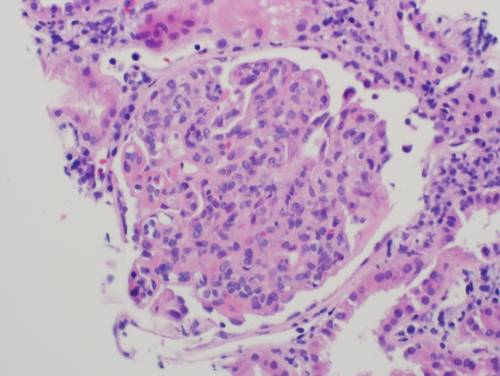
Repeat biopsy shows:


Which of the following is the most appropriate therapy for her current condition?


A. Mycophenolate and steroid
B. Mycophenolate, tacrolimus and steroids 
C. Rituximab 
D. I.V Immunoglobulins 
E. Plasma exchange  
Omitted
The correct answer is B

Mycophenolate, tacrolimus and steroids 

Explanation: 

This patient had membranous lupus being treated with Euro-Lupus regime. Activity in the urine with a positive ANA and dsDNA and low C3, C4 suggests activity of the disease. The biopsy is showing active lupus nephritis combined with features of class IV & V diffuse proliferative GN. Patients with both DPGN and membranous lesions are more likely to be resistant to standard induction regimens with cyclophosphamide or MMF with steroids. In these patients, combined treatment with MMF and tacrolimus is recommended. This was suggested by Bao H. et al in a small, short term prospective trial in which 40 patients with diffuse proliferative plus membranous lupus nephritis were randomly assigned to induction therapy with MMF (0.75g to 1g/d) and tacrolimus (3-4 mg/d) or intravenous cyclophosphamide alone .All patients received steroids. At nine months there was significantly higher rate of complete remission in patients treated with MMF and tacrolimus as compared to cyclophosphamide (65% versus 15%).

(Choice A) Mycophenolate and steroids can be used in patients who have received cyclophosphamide and are resistant to it.

(Choice C and D) Rituximab and I.V. Immunoglobulins can be used in patients with lupus nephritis who have failed to respond to cyclophosphamide, MMF and steroids.

(Choice E) Plasma exchange is recommended in patients with SLE and (TTP). 

KDIGO Clinical Practice Guidelines for Glomerulonephritis recommends the following treatment for resistant lupus nephritis:

Treatment of resistant disease 12.9.1: In patients with worsening S. Cr and/or proteinuria after completing one of the initial treatment regimens, consider performing a repeat kidney biopsy to distinguish active LN from scarring. (Not Graded) 12.9.2: Treat patients with worsening S. Cr and/or proteinuria who continue to have active LN on biopsy with one of the alternative initial treatment regimens. If patient has received cyclophosphamide use MMF and if patient has received MMF use cyclophosphamide (Not Graded) 12.9.3 

KDIGO suggests that non -responders who have failed more than one of the recommended initial regimens (cyclophosphamide, MMF or CNI) may be considered for treatment with rituximab, IV Immunoglobulins, or CNIs. (2D).


Copyright © ABIM Exam World
Created On: 09/20/2017
Last Modified: 08/06/2018

End

Previous Next
Multiple
  • Test Id: 19515678623c961dcb
  • QId: 165260
  • 7 of 39
  • Notes
  • Calculator
  • Feedback

You are rounding on your patients in the dialysis unit and seeing a 65-year-old gentleman with ESRD due to chronic interstitial disease. He also has a history of diet-controlled diabetes mellitus and hypertension. His other past medical history is significant for dyslipidemia, coronary artery disease, hypothyroidism, gout and depression. He has been hospitalized in the recent past for swelling and pain of his right great toe. He was seen by the foot doctor, a scan was done and eventually the great toe had to be amputated.  He has been on hemodialysis 3 times a week. His weekly Kt/V is 1.9. You are conducting the monthly blood work review for this patient. You note that his hemoglobin has been persistently low for past few monthly blood draws. He is currently on 100 mcg of Darbepoetin weekly on dialysis. On enquiry there is no history of blood loss in the form of hematemesis, melena, hematochezia or hemoptysis. His active medication list includes Losartan, Atorvastatin, Calcitriol, multivitamin supplements, paroxetine, allopurinol, aspirin.

His pertinent blood work is as follows:

Test

Result

WBC

4500 cells /cumm

Hemoglobin

8.2 g/dL

Platelet count

450 thousand /cumm

Reticulocyte count

Normal

Iron

55 (range 50-150)

Total iron binding capacity

250 g/dl (range 250-310)

Transferrin saturation

20%

Ferritin

1400 ng/ml (range 20-235)

Haptoglobin

400 mg/dl (range 83-267)

Lactate dehydrogenase

240 U/L (range 80-225)

Total bilirubin

1.0 mg/dl

Folate

7 ng/ml (range 1.8-9.0)

B12

500 pg/ml (range 200-800)


Peripheral blood smear.

Normal RBC morphology, few burr cells.


Which of the following is true about this patient’s anemia?


Omitted

Correct Answer: Option C: This patient has chronically inflamed state which is contributing to his anemia.


Explanation:

10-15% of patients who have been receiving erythrocyte estimating agents (ESA) develop resistance. There are multiple reasons why ESRD patients develop resistance.


ESA resistance occurs due to the following reasons:

  1. Iron deficiency.

  2. Chronic inflammation.

  3. Under-dialysis.

  4. Hemolysis.

  5. Folate and B12 deficiency.

  6. Chronic blood loss.

  7. Anti EPO antibodies.

  8. Pure red cell aplasia.

  9. Failed chronic renal allograft.

  10. ACEI/ARB.

  11. Aluminum overload.

  12. Hyperparathyroidism.

  13. Hematological disorders or malignancy.


Option A: Incorrect option. ESRD is associated with erythropoietin deficiency. Patient has been initiated on ESA already. There is no point in measuring EPO levels. There is no evidence of measuring EPO levels in management of anemia in CKD.

Option B: Incorrect option. ESRD is an inflamed state. In inflammatory milieu there is increased production of Hepcidin. The hepatic iron-regulatory hormone Hepcidin and its receptor, the cellular iron exporter Ferroportin, constitute a feedback-regulated mechanism that maintains adequate plasma concentrations of iron-transferrin for erythropoiesis and other functions, ensures sufficient iron stores, and avoids iron toxicity. In chronic kidney disease, inflammation and impaired renal clearance increases plasma hepcidin, inhibiting duodenal iron absorption and sequestering iron in macrophages. These effects of hepcidin can cause systemic iron deficiency, decreased availability of iron for erythropoiesis, and resistance to endogenous and exogenous erythropoietin.

Choice C: Correct option. Refer explanation for option B.  He had pain, swelling of his right great toe, a foot doctor sees him, a bone scan is done and subsequently the amputation. All suggestive of an infective etiology probably osteomyelitis.There is a temporal relationship between patients’ anemia and underlying chronic inflammatory state.

The high ferritin is also suggestive of inflamed state.

Choice D: Incorrect option. Pure red cell aplasia, a form of severe ESA hypo-responsiveness mediated by anti-erythropoietin antibodies, was first reported with certain formulations of Epoetin alfa but has now been reported with all commercially available forms of ESA. This syndrome presents with rapid onset of severe anemia (hemoglobin <7 g/dl), severe reticulocytopenia (reticulocyte count <10,000/?l) and marked elevations in serum ferritin level (>1000 ng/ml) and transferrin saturation (>70%) resulting from low iron utilization. Pure red cell aplasia is unlikely given the absence of characteristic laboratory findings. Moreover, the patient did not receive Epoetin alfa.


Choice E: Incorrect option. Under-dialysis leads to anemia due the same mechanism mentioned earlier in option B. Under-dialysis worsens the uremic milieu which in turn leads to inflammatory state. This leads to anemia. Patient in this clinical vignette has been dialysed appropriately. His weekly Kt/V is 1.9, which is above the target goal of 1.7

Copyright © ABIM Exam World
Created On: 10/22/2018
Last Modified: 04/17/2021

End

Previous Next
Multiple
  • Test Id: 19515678623c961dcb
  • QId: 165261
  • 8 of 39
  • Notes
  • Calculator
  • Feedback

50-year-old female patient whos group B is being evaluated for kidney transplant surgery. She had ESRD secondary to analgesic nephropathy and is on hemodialysis for last 5 years. She has had multiple sensitization events in the form of 3 pregnancies and several blood transfusions. Her current calculated PRA against class I antigen is 97% and against class II antigen is 99%. She has been enrolled in the national highly sensitized recipient program.

Her husband who is blood group matched came forward as a potential kidney donor but she had positive Flow B and T Cell Cross match against him. Single antigen bead assay demonstrated that she has donor specific antibodies against class II across DQB*15 and DPB*14. This transplant did not materialize as patient declined desensitization protocol. Now her younger brother comes forward as a potential donor. He is blood group A and the flow B and T cell cross match is negative with no demonstrable donor specific antibodies against this donor.Patient wants to know more about ABO incompatible transplant.


Which of the following statements about the ABO incompatible transplant is correct?


A. Three-year graft survival is inferior to blood group compatible transplants.
B. The infectious and bleeding complications post ABOI kidney transplant are the same as blood matched kidney transplant.
C. All patients undergoing ABOI transplant need to undergo desensitization using IVIg, Plasma exchange, Rituximab irrespective of their donor/recipient pair Anti ABO titers for optimal outcomes.
D. C4d staining on protocol biopsies is common feature and does not necessarily mean an antibody mediated rejection process in the absence of allograft dysfunction.
Omitted

Correct answer: Option D: C4d staining on protocol biopsies is common feature and does not necessarily mean an antibody mediated rejection process.


Explanation:


Choice A: Three-year graft survival is inferior to blood group compatible transplants is incorrect A comprehensive database analysis of 1420 ABOI living donor (LD) kidney transplants performed in 101 centers from 2005 to 2012 compared graft and patient survival to a matched cohort of ABO-compatible transplant recipients. Three-year graft and patient survival were ultimately identical. 1


Choice B: The infectious and bleeding complications post ABOI kidney transplant as same as blood matched kidney transplant is also incorrect. Using USRDS and Medicare data from 2000–2007, 119 ABOI (non-A2 donor) transplant recipients were identified. Compared with ABO-compatible recipients, the risks of infectious and hemorrhagic complications were significantly higher, with a 2.2-fold higher risk of pneumonia, a 3.5-fold higher risk of wound infections, a 56% higher risk of pyelonephritis, and a nearly 2- fold higher risk of hemorrhage 2


Choice C: All patients undergoing ABOI transplant need to undergo desensitization using IVIg, Plasma exchange, Rituximab irrespective of their donor/recipient pair Anti ABO titers for optimal outcomes is also an incorrect answer. Historically, ABOI transplantation has been successful when performed after desensitization with plasmapheresis, intravenous Ig (IVIG), rituximab, and/or splenectomy to achieve ABO IgG antibody titers 1:4. A recent publication demonstrated that these intensified treatments might not be necessary in donor/recipient pairs who have low-moderate titer ABO incompatibility 3


Choice D: C4d staining on protocol biopsies is common feature and does not necessarily mean an antibody mediated rejection process in the absence of allograft dysfunction is the correct answer C4d staining is not an uncommon feature seen in the protocol biopsies done in ABOI kidney transplant recipients. In the absence of allograft dysfunction, the C4d staining has no clinical relevance and is just a part of the graft accommodation.

Copyright © ABIM Exam World
Created On: 10/30/2018
Last Modified: 10/23/2020

End

Previous Next
Multiple
  • Test Id: 19515678623c961dcb
  • QId: 165263
  • 9 of 39
  • Notes
  • Calculator
  • Feedback

A 19-year-old woman, African American descent, comes to clinic for follow up visit. She has been found to have type 1 diabetes mellitus since the age of 12 years of age. She has been using insulin pump for the last 5 years. She reports no hypoglycemic symptoms and has been monitoring blood sugar using flash glucose monitor. She reports infrequent hypoglycemic episodes all being self-managed. She met with an ophthalmologist for eye screening and has no retinopathy. She exercises regularly for 30 mins. Her vitals recording shows BP of 127/66 mmHg. Her BMI is 22.2.  Systemic  examination is unremarkable. 

Her laboratory investigation is as follows.


Characteristic

value

Hemoglobin

13.2 gm/L 

WBC count

7.8 X 10³/cubic mm

Platelet count

241 X 103/cubic mm

Segmented Neutrophils 

Lymphocytes

Monocytes

Band neutrophils 

Eosinophils

Basophils

60%

36%

2%

0%

2%

0%

Sr. Sodium

136 mEq/L

Sr. Potassium

4.2 mEq/L

Sr. Creatinine

0.6 mg/dL

eGFR using CKD-EPI

153.1 ml/min/1.73m2

Sr. Bicarbonate

24 mEq/L

Sr. Chloride

101 mEq/L

Total Bilirubin

1.0 mg /dL

AST

16 U/L

ALT

18 U/L

Sr. Albumin

4.0 g/dL

HBA1C

8.2%

Sr. Calcium

10 mg/dL

Urine dipstick

pH- 5.4

Albumin-nil

 no blood

 no WBCs

24-hour urinary protein 

86 milligrams/day


What is the MOST LIKELY False statement regarding renal hyper filtration stage of Diabetic Kidney Disease in this patient?

A. Renal hyper filtration is attenuated by SGLT2 inhibition.
B. Renal hyper filtration occurs in both type 1 and type 2 diabetes mellitus.
C. Obesity can also lead to single-nephron hyper filtration.
D. Incretins like GLP-1 and GIP are neutral in terms of altering renal hemodynamics unlike SGLT2 blockers.
Omitted

The Correct Answer is Option D : Incretins like GLP-1 and GIP are neutral in terms of altering renal hemodynamics unlike SGLT2 blockers.


Supra-physiologic elevation in GFR is observed early in the natural history of type 1 and type 2 diabetes mellitus which is due to glomerular hyperfiltration. Pathogenesis of hyper filtration in diabetes is complex with a prominent role for hyperglycemia and distorted insulin levels especially in early diabetes and pre-diabetes.Dilatation of the afferent (pre-capillary) glomerular arteriole plays an important role in the hyper-filtration response, by raising both the intra-glomerular pressure and renal blood flow.


The effect of incretins can be demonstrated by experiment using GLP-1 receptor agonists (GLP-1RA) and dipeptidyl peptidase (DPP)–4 inhibitors which are associated with renal hemodynamic effects, potentially beyond glycemic control. These observations have been attributed to a GLP-1–mediated inhibition of NHE3 (which assembles with DPP-4 in the proximal tubular brush border), thereby reducing proximal sodium reabsorption and GFR through activation of TGF (tubuloglomerular feedback).


Option A :  In an 8-week study, empagliflozin in T1DM patients with whole-kidney hyper filtration (mean GFR 172±23 ml/min per 1.73 m2) demonstrated a glucose-independent 19%decrease in GFR, which was associated with a decline in ERPF (estimated renal plasma flow) and estimated glomerular pressure and increase in afferent arteriolar resistance, as assessed by the Gomez equations. SGLT2 inhibition could reduce (single-nephron) hyperfiltration in diabetes by restoring sodium-chloride concentration at the macula densa and subsequent TGF mediated afferent arteriolar vasoconstriction.


Option B : Reported prevalence of hyper filtration at the whole-kidney level vary greatly: between 10% and 67% in type 1 diabetes mellitus (T1DM) (with GFR values up to 162 ml/min per 1.73 m2), and 6%–73% in patients with type 2 diabetes (T2DM) (up to 166 ml/min per 1.73 sq. m. 


Option C: Independent of diabetes and glucose levels, body weight also augments GFR (by about 15% in obese to about 56% in severely obese non-diabetic subjects).

Copyright © ABIM Exam World
Created On: 10/31/2018
Last Modified: 10/23/2020

End

Previous Next
Multiple
  • Test Id: 19515678623c961dcb
  • QId: 165264
  • 10 of 39
  • Notes
  • Calculator
  • Feedback

A 68-year-old gentleman, Caucasian descent, comes to clinic for follow up visit. He is known to have type 2 diabetes mellitus for the past 18 years. His father had diabetes from 40 years of age and developed kidney disease requiring dialysis after 15 years of diabetes. He reports no symptoms. He has been having hypertension and coronary artery disease with history of  PCI 2 years ago. He has non-proliferative diabetic retinopathy. His medications are sitagliptin, gliclazide and metformin in addition to losartan and hydrochlorothiazide. He has been monitoring blood sugar at home and reports no hypoglycemia. He exercises at least at least 30 minutes per day. His vitals recording shows BP of 168/66 mm Hg. His BMI is 29.2.  Systemic  examination is unremarkable.

His laboratory investigation is reported as follows.

Characteristic

value

Hemoglobin

12.2 gm/L

WBC count

6.8 X 10³/cubic mm

Platelet count

241 X 103/cubic mm

Segmented Neutrophils

Lymphocytes

Monocytes

Band neutrophils

Eosinophils

Basophils

60%

36%

2%

0%

2%

0%

Sr. Sodium

139 mEq/L

Sr. Potassium

4.9 mEq/L

Sr. Creatinine

1.2 mg/dL

Sr. Bicarbonate

22 mEq/L

Sr. Chloride

101 mEq/L

Total Bilirubin

1.0 mg /dL

AST

16 U/L

ALT

18 U/L

Sr. Albumin

4.0 g/dL

HBA1C

7.8%

Sr. Calcium

10 mg/dL

Urine dipstick

pH- 5.4

Albumin-trace

 no blood

 no WBCs

24-hour urinary albumin

200 milligrams/day


What is the MOST LIKELY correct statement regarding clinical diagnosis of Diabetic Kidney Disease in this patient ?

A. Diabetic Kidney Disease previously called as diabetic nephropathy can be diagnosed clinically with renal biopsy only.  
B. Presence of microalbuminuria is adequate for clinical diagnosis of Diabetic Kidney Disease. 
C. Presence of hematuria without non-diabetic kidney disease is impossible in Diabetic Kidney Disease as diabetic kidney disease is a non-proliferative glomerular disease. 
D. Family history of Diabetic Kidney Disease is associated with renal involvement in Diabetes.
Correct

The Correct Answer is Option D: Family history of Diabetic Kidney Disease is associated with renal involvement in Diabetes.

 Explanation:

Familial studies have demonstrated clustering of diabetic nephropathy. Patients with DM with a first-degree relative with T1/T2DM and diabetic nephropathy have more risk for developing diabetic nephropathy than those without an affected relative. This familial clustering has also been well documented in the Pima Indian population. The candidate genes identified are glucose transporter 2(GLUT2), transforming growth factor beta (TGF- ?), and endothelial nitric oxide synthase (eNOS). 

Option A:  Diabetic nephropathy is a clinical syndrome characterized by the following:

·         Persistent albuminuria (>300 mg/d) that is confirmed on at least 2 occasions 3-6 months apart

·         Progressive decline in the glomerular filtration rate (GFR)

·         Elevated arterial blood pressure 

 Hence kidney biopsy is not a mandatory investigation to diagnose diabetic kidney disease.

 Option B:  If the amount of urine albumin exceeds 30 mg/d and is less than 300 mg/d it is called microalbuminuria, and if it is greater than 300 mg/d it is called macro albuminuria or overt albuminuria. Microalbuminuria is present in 5-7% of normal individuals and is associated with cardiovascular mortality and morbidity. It is marker of endothelial dysfunction in type 2 diabetes mellitus. Presence of microalbuminuria alone with diabetes cannot be clinically diagnostic of diabetic kidney disease.

Option C:  Micro hematuria has been demonstrated in biopsy studies with isolated diabetic nephropathy. Red blood cell casts have also been described in patients with diabetic nephropathy. However, it is important to rule out other glomerular and extra-glomerular causes of hematuria.

Copyright © ABIM Exam World
Created On: 10/31/2018

End

Previous Next
Multiple
  • Test Id: 19515678623c961dcb
  • QId: 167322
  • 11 of 39
  • Notes
  • Calculator
  • Feedback

A 45-year-old female is wait listed for kidney transplant. Her native kidney disease is IgA nephropathy. Her blood group is B and her CPRA is 90%. You are seeing her in the clinic as a part for kidney transplant work. As a part of documentation, you discuss with her about Public Health Service – Increased Risk Donors (PHS-IRD). You explain to her about PHS-IRD and if she would consent for it. Your rational for explaining this is a high wait time for blood group B in your allocation area especially in the setting of CPRA 90%. Which of the following statements regarding the PHS-IRD is TRUE ?


A. As per the new guidelines there is no need to take an informed consent for PHS-IRD from the potential recipient. 
B. PHS-IRD kidneys have a discard rate of 2.5-fold. 
C. The kidney from PHS-IRD have an infection transmission risk anywhere between 10-15% depending on the type of infection. 
D. Recipient who received IRD kidneys have increased risk of death at 6 months compared to those who declined the offer.
E. There is increased risk of allograft loss in recipients of PHS-IRD kidneys. 
Correct

Correct Answer. Option B. PHS-IRD kidneys have a discard rate of 2.5-fold.


Explanation. 

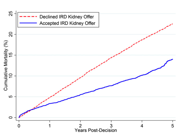
The United States Public Health Service (PHS) redefined donors who were previously classified by the Centers for Disease Control at increased risk for transmission of hepatitis B virus (HBV), hepatitis C virus (HCV), and HIV. The number of deceased donors that are part of this classification has increased dramatically because of the opioid epidemic, comprising over 20% of donor kidneys. Overall, overdose-death donors accounted for 1.1% of organ donors in 2000 and 13.4% in 2017. Importantly, transplantation candidates and providers should be well versed in the very low risk of disease transmission from these donors, all significantly ,1% even under the highest-risk circumstances (intravenous drug overdose, syringe-on-person). Unfortunately, “PHS increased-risk donor” (IRD) status is independently associated with a nearly 2.5-fold increased odds of turndown. An analysis by Bowring et al. used SRTR data from 104,998 kidney transplantation candidates who were offered IRD kidneys that were eventually accepted. The median KDPI of these kidneys was 30 (interquartile range, 16–49). Importantly, after 5 years, only 31.0% of candidates who declined IRDs received non-IRD DDKTs later; the median KDPI of these non-IRD kidneys was 52. Those who accepted an IRD had a substantially lower risk of death at 1 to 6 months after decision (aHR, 0.50; 95% CI, 0.67 to 0.90; P=0.006) and beyond 6 months after decision (aHR, 0.46; 95% CI, 0.52 to 0.58; P< 0.001). A single-center report of PHS-IRD kidney utilization reviewed offers made to 2423 kidney transplant candidates from June 2004 to May 2005; 1502 ultimately received a transplant with or without a PHS-IRD kidney. Acceptance of a PHS-IRD kidney offer was associated with lower risk of mortality (3.63% versus 11.6%; aHR, 0.467; P = 0.0008) and decreased risk of allograft loss compared with non– PHS-IRD recipients (P=0.007), with no transmission of HCV, HBV, or HIV.


Copyright © ABIM Exam World
Created On: 05/12/2020
Last Modified: 01/28/2021

End

Previous Next
Multiple
  • Test Id: 19515678623c961dcb
  • QId: 167339
  • 12 of 39
  • Notes
  • Calculator
  • Feedback

All the following drugs mentioned below cause hyperkalemia. All the drugs act by a specific mechanism to induce hyperkalemia. Only one drug acts by a different mechanism of action. Which is the drug which induces hyperkalemia via a different mechanism than others?  


A. Heparin.
B. Low molecular weight heparin. 
C. Aminoglutethimide. 
D. Dabigatran.
E. Calcineurin inhibitors. 
Incorrect

Correct Answer. Option D. Calcineurin inhibitors. 


Explanation. 

Heparin, Low molecular weight heparin, Aminoglutethimide and Dabigatran all cause impaired adrenal hormone metabolism. These drugs cause potent inhibition of adrenal hormone synthesis leading to hyperkalemia. 

The hyperkalemia seen with calcineurin inhibition is likely multifactorial and relates to inhibitory effects on Na+-K+-ATPase in collecting ducts and possibly to distal tubular acidosis. In addition, there is evidence that decreased numbers of mineralocorticoid receptors, which are detected in 75% of patients who are treated with cyclosporine, lead to hyperkalemia and metabolic acidosis as a result of aldosterone resistance. Recently, it was demonstrated that cyclosporine reduces paracellin-1 expression in thick ascending limb cells. The resulting decrease in magnesium transport likely contributes to the magnesium wasting and hypomagnesemia induced by cyclosporine, which is associated with chronic interstitial fibrosis, a faster rate of decline of kidney function, and increased rates of graft loss in renal transplant recipients with CNI nephrotoxicity. Finally, it was shown that cyclosporine indirectly opens ATP-sensitive K+ channels by inhibition of calcineurin, which could contribute to the CNI-associated hyperkalemia. 

Copyright © ABIM Exam World
Created On: 05/18/2020
Last Modified: 01/28/2021

End

Previous Next
Multiple
  • Test Id: 19515678623c961dcb
  • QId: 167340
  • 13 of 39
  • Notes
  • Calculator
  • Feedback

A 25-year-old man is referred to you for evaluation of nephrotic syndrome. A kidney biopsy is done, and it shows minimal change disease. His BP is 150/90 mmHg with a heart rate of 70 bpm. His current medications are as follows: Prednisone 40 mg once daily, furosemide 40 mg twice daily, Ramipril 10 mg once daily and Calcium + vitamin D supplementation. Clinical exam shows raised JVD, 3 + pitting edema till the ide thigh. His blood work shows Sr creatinine of 0.9 mg/dL, BUN 35 mg/dL and his Sr potassium is 3.2 mg/DL. Urine analysis is bland and his 24 hr urine protein is 5.8g. He is following his salt restriction strictly. After a month’s follow up his proteinuria has reduced to 4.0 g/24 hrs but his edema feet has unchanged. His Sr albumin has improved from 1.9 g/L to 2.8 g/L. Addition of which of the following drugs will benefit the patient? 

A. Metolazone.
B. Amiloride. 
C. Chlorthalidone. 
D. Cyclophosphamide. 
E. Amlodipine. 
F. Losartan. 
Omitted

Correct Answer. Option B. Amiloride. 


Explanation. 


Two major factors, both of which lead to retention, have been thought to be responsible for the development of edema in patients with the nephrotic syndrome; it is likely that both contribute to a variable degree in individual patients:

  1. Primary sodium retention that is directly induced by the renal disease (overfill hypothesis).
  2. Secondary sodium retention in which the low plasma oncotic pressure due to hypoalbuminemia promotes the movement of fluid from the vascular space into the interstitium, leading to underfilling of the vasculature and activation of the renin-angiotensin-aldosterone system (underfill hypothesis).

The clinical importance of distinguishing between these mechanisms is the ability to tolerate diuretic therapy. Diuretics are well tolerated in patients with renal sodium retention but, if underfilling is the primary mechanism, can lead to worsening hypovolemia as evidenced clinically by an elevation in serum creatinine. 


Studies in experimental animals with unilateral nephrotic syndrome or glomerulonephritis suggest that primary sodium retention in these disorders is due to increased sodium reabsorption in the collecting tubules, which is also the site of action of atrial natriuretic peptide (ANP) and the related renal hormone urodilatin. This has been called the overfill hypothesis since primary renal sodium retention leads to volume expansion


Increased activity of the epithelial sodium channel (ENaC) may contribute to sodium retention. Serine proteases are aberrantly filtered in the nephrotic syndrome, leading to increased concentration in the urine. Plasminogen (which is activated by epithelial urokinase-type plasminogen activator [uPA] to plasmin) in nephrotic urine may activate ENaC via proteolytic cleavage of the gamma chain, providing a potential mechanism by which filtered proteins cause sodium retention. It also explains the observation that remission from the nephrotic syndrome is generally preceded by a decrease in urinary protein excretion. In an animal model of nephrotic syndrome, mice treated with the serine protease inhibitor aprotinin normalized urinary serine protease activity and prevented sodium retention. A similar result was seen when rats with nephrosis were treated with the ENaC inhibitor amiloride.

Hence Amiloride is the best answer for this clinical vignette. 

Copyright © ABIM Exam World
Created On: 05/18/2020
Last Modified: 01/28/2021

End

Previous Next
Multiple
  • Test Id: 19515678623c961dcb
  • QId: 167322
  • 14 of 39
  • Notes
  • Calculator
  • Feedback

A 15 year-old boy is brought to the ER by his foster mother who states that when she got home from work she noticed he was acting very strange. He had slurred speech and seemed confused. He appeared to be very uncoordinated and she was not sure if he fell or hit his head. She states that he is somewhat a troubled boy but doesn’t know much about his history as he has been in and out of the foster care system out of state. On physical exam, he is tachycardic and has tachypnoea. Pupils are dilated, but there is no nystagmus. A fundoscopic exam shows hyperemia of the optic disk. He is relatively uncooperative but not aggressive or hostile. When asked about suicidal thoughts he responds only with inaudible mumbling. His foster mother left for work 10 hours prior and assumed he left for school. She is not sure when these symptoms began or what may have initiated them. P is 105/ min, BP is 140/90 mm Hg, RR is 28/min, and T is 97.1 F. Laboratory examination is as follows: 

Na   135 mEq/L                                            

K   5.0 mEq/L

CL   105 mEq/L

BUN  19 mg/dL

Cr   1.3 mg/dL         

HCO3  8 mEq/L  

Glucose  100 mg/dL         

pH   7.3          

pO2   90 mmHg

pCO2  22 mmHg

Measured serum osmolarity  320 mmol/L

What is the next step in management?

A.  Gastric lavage
B.  N-acetylcystiene and activated charcoal
C. Fomepizole 
D. Fomepizole and Hemodialysis 
E.  Obtain serum levels of salycylate, methanol and ethylene glycol levels
Correct
The correct answer is D
Hemodialysis and fomepizole

Explanation: 
While this patient's case may appear to be vague, there are three strong indicators for the most likely diagnosis:
1)    This patient has metabolic acidosis with profound Osmolar gap, which is a strong indicator of volatile alcohol related products. 
2)    Hyperemia of the optic disk (or other ocular symptoms) in addition to profound anion gap should make methanol toxicity a top differential diagnosis.  
3)    In addition to the serum anion gap, there is a serum Osmolar gap. (Measured Osm – Calculated Osm) where:

Calculated Osmolarity = 2(Na) + BUN/2.8 + Glu/18

***Anytime there is an osmolar gap in metabolic acidosis, your differentials can be narrowed down to volatile alcohol related substances. ***  --------------->    IMPORTANT ABIM BOARD POINT

The best treatment for methanol and ethylene glycol is to remove the toxic byproducts from circulation. The definitive therapy is Hemodialysis. Fomepizole is given to block alcohol dehydrogenase, thereby stopping the formation of toxic byproducts. However, it does not remove them from circulation, only dialysis can do this.

(Choice A) Gastric lavage is almost never the correct answer, especially with ingestion of an unknown substance. Caustic substances can cause further damage on the way out with the use of gastric lavage. Esophageal perforation is potentially a fatal risk.

(Choice B) N-acetyl cystiene is the treatment for acetaminophen toxicity however timing is important and drug levels should be drawn first to determine the need for treatment. If ingestion occurred more than 24 hours earlier, treatment has no effect and is therefore not given.

(Choice C, D) Refer to the explanation section above.

(Choice E) When suspicion for methanol poisoning is high, treatment should not wait for diagnosis confirmation. The presence of ocular involvement is an ominous sign, and the risk of blindness is increased. The benefits outweigh the risk of treating before confirming diagnosis in this case.


Copyright © ABIM Exam World
Created On: 09/13/2017
Last Modified: 12/30/2017

End

Previous Next
Multiple
  • Test Id: 19515678623c961dcb
  • QId: 167339
  • 15 of 39
  • Notes
  • Calculator
  • Feedback

All of the following are helpful in predicting AV Graft stenosis EXCEPT:

A. Hyperpulsatility of the AV Graft
B. Ruduced pulse augmentation
C. Increased bleeding and clots 
D. Decreased KT/V
E. Decreased Blood Flow rate
F. Surveillance of the graft
Incorrect
The correct answer is F

Surveillance of the graft

Explanation:

Intra-graft or venous outflow stenosis – A strong pulse in the AV graft - HYPERPULSATILITY suggests an increase in resistance as occurs with a venous stenotic lesion. The intensity of this pulse is directly proportional to the severity of the stenosis. For this reason, in an AV graft Hyperpulsatility can be considered as an indicator of impending AV graft stenosis.

Arterial stenosis – REDUCED PULSE AUGMENTATION suggests inflow stenosis due to stenosis of the arterial anastomosis or of the feeding artery. Although pulse augmentation is commonly performed, it is less sensitive for the detection of inflow stenosis in AV grafts compared with AV fistulas.

INCREASED BLOOD CLOTS, DECREASED KT/V, and DECREASED BLOOD FLOW RATE are all suggestive of impending AV graft stenosis. Often patients are seen with dilated, collateral veins over the arm and chest wall (very important to examine your hemodialysis patients after taking off their shirts) suggestive of central venous stenosis - The classic physical finding in a patient with a significant central venous stenosis is diffuse upper extremity edema. Subcutaneous collateral veins are frequently evident over the chest. Swelling and collateral veins are caused by generalized venous hypertension of the extremity, which occurs in central, but is rare with peripheral lesions. 

SURVEILLANCE  does not predict AV Graft stenosis. It is not a sensitive or specific modality to detect the same accurately.

Copyright © ABIM Exam World
Created On: 09/14/2017
Last Modified: 08/06/2018

End

Previous Next
Multiple
  • Test Id: 19515678623c961dcb
  • QId: 167340
  • 16 of 39
  • Notes
  • Calculator
  • Feedback

A 40 year-old pleasant African man with ESRD secondary to FSGS started automated peritoneal dialysis. His prescription includes 2.5 L and 3 exchanges over 8 hours at night with a last fill of 2 L. He has a urine output of 1000 mL/day. A typical ultrafiltration on cycler is used at 1000 mL. Average drain volume of the day dwell was 1500 mL prior to going on the cycler at night. 

He came with complains of lower abdominal wall edema extending to the scrotum over the past 5 days. Without any change in the dialysis prescription, his drain volume before going on the cycler dropped to 900 mL, and the ultrafiltration volume on the cycler came down to 100 mL. He reports no pain with fill or drain. 

What is the next step?


A. Chest X ray PA and lateral view 
B. Drain the fluid middle of the day to reduce the dwell time
C. PD catheter manipulation
D. Abdominal CT scan with contrast in the dialysate
E. Switch to hemodialysis 
F. Pleurodesis 
Omitted
The correct answer is D 

Abdominal CT scan with contrast in the dialysate

Explanation:

Typical ultrafiltration failure from peritoneal membrane failure presents gradually. More frequently, we see an apparent ultrafiltration failure from other reasons. 

Catheter malfunction can present with problem with inflow, outflow or both. A kink in the catheter poses problem with inflow and outflow. 

Omentum or blood clots clogging the catheter can cause only outflow problem. Fibrin clots, constipation with loaded rectum, and displaced PD catheter also cause outflow obstruction. X ray KUB is very helpful in diagnosing displaced catheter.   

FLUID LEAK leak into the abdominal wall causes swelling in the lower abdomen extending down to the scrotum or mons pubis and decrease in the drain volume. Usually this can be treated by temporarily doing low volume PD or transferring to HD for about 6 weeks. Such leaks starting few years after initiating PD usually do not respond to this approach and often requires placement of a new PD catheter.

A patent tunica vaginalis testes will cause UNILATERAL OR BILATERAL SCROTAL SWELLING without much swelling of the abdominal wall sometimes associated with decrease in the drain volume. A CT scan of the abdomen with ‘IV’ contrast into the peritoneal fluid can help in diagnosing the leak into the abdominal wall and scrotal leak. Patent tunica vaginalis testes requires surgery. Generally hemodialysis is not required after the surgery and low volume peritoneal dialysis can be resumed. This patient presents with signs of leak of fluid into the abdominal wall. A chest X ray is not required, so (Choice A) is wrong.

Draining during the middle of the day helps if the membrane is very permeable with very high D/P creatinine ratio on PET. This does not help in abdominal wall leak of dialysate. Therefore, (Choice B) is also wrong.

PD catheter manipulation (Choice C) is wrong because this patient does not have displaced PD catheter from the information given.

(Choice D) is the correct answer. If the clinical presentation is very convincing, most patients are treated with reducing the dialysate volume and if possible keeping the abdomen dry during the day. Sometimes these patients need to be switched to hemodialysis for 4-6 weeks before PD is resumed. Hence temporary switch to HD would be a correct answer, but unqualified switch to HD as is stated in answer E would be a wrong choice. Therefore, (Choice E) is the wrong or less appropriate answer.

ABDOMINAL ADHESIONS cause decrease in the drain volume as well and require surgical treatment with low volume PD or temporary HD after that.

DIAPHRAGMATIC LEAK into the pleural space presents with cough and shortness of breath without peripheral signs of fluid overload few weeks after starting peritoneal dialysis (not the presentation in this patient). Usually, such patients present with right sided pleural effusion clinically and on chest X ray PA and lateral view. These patients can be treated with temporary transfer to HD, pleuridesis and return back to PD 4-6 weeks later. Permanent switch to hemoidialysis without pleurodesis is another option.


TREATMENT OPTIONS OF VARIOUS ABDOMINAL/CHEST PROBLEMS AS MENTIONED ABOVE IN PATIENTS ON PERITONEAL DIALYSIS IS FREQUENTLY TESTED ON BOARDS. PLEASE KNOW THE TREATMENT OPTIONS.


Copyright © ABIM Exam World
Created On: 09/14/2017
Last Modified: 08/06/2018

End

Previous Next
Multiple
  • Test Id: 19515678623c961dcb
  • QId: 165212
  • 17 of 39
  • Notes
  • Calculator
  • Feedback

The ACCOMPLISH trial is the first major trial addressing the issue of combination therapy in 11,506 patients who were at high cardiovascular risk. The goal blood pressure was less than 130/80 mm Hg in the patients with diabetes or impaired renal function, and less than 140/90 mm Hg in the patients with prior cardiovascular disease.

Which of the following combinations of blood pressure medications was the best in reducing cardiovascular events and slowing the progression of nephropathy in patients with hypertension who were at high risk for such events?

A. ACEI + Diuretics
B. ACEI + CCB 
C. ACEI + Beta-blocker 
D. CCB + Beta-blocker 
Incorrect
The correct answer is B
ACEI + CCB

Explanation:

• The Avoiding Cardiovascular Events through Combination Therapy in Patients Living with Systolic Hypertension  - (ACCOMPLISH) trial showed that initial antihypertensive therapy with benazepril plus amlodipine was superior to benazepril plus hydrochlorothiazide in reducing cardiovascular morbidity and mortality.

• ACCOMPLISH ACCOMPLISH was a double-blind, randomized trial undertaken in five countries (USA, Sweden, Norway, Denmark, and Finland). 11 506 patients with hypertension who were at high risk for cardiovascular events were randomly assigned via a central, telephone-based interactive voice response system in a 1:1 ratio to receive benazepril (20 mg) plus amlodipine (5 mg; n=5744) or benazepril (20 mg) plus hydrochlorothiazide (12.5 mg; n=5762), orally once daily. Drug doses were force-titrated for patients to attain recommended blood pressure goals.

• The primary end point was the composite of death from cardiovascular causes, nonfatal myocardial infarction, nonfatal stroke, hospitalization for angina, resuscitation after sudden cardiac arrest, and coronary revascularization.

• The benazepril-amlodipine combination was superior to the benazepril-hydrochlorothiazide combination in reducing cardiovascular events in patients with hypertension who were at high risk for such events.

Copyright © ABIM Exam World
Created On: 09/20/2017
Last Modified: 01/25/2021

End

Previous Next
Multiple
  • Test Id: 19515678623c961dcb
  • QId: 165246
  • 18 of 39
  • Notes
  • Calculator
  • Feedback

A 32 year-old male is brought to renal clinic with history of hematuria, oedema feet, and puffiness of face. He gives a history of fever and sore throat a week ago. He also complains of breathlessness on exertion and oliguria. Physical examination shows: Pulse 100/min, BP 150/100 mm Hg, and Temp. 37.4 C. He is pale. He has puffiness of face and oedema feet. Systemic examination-unremarkable. Laboratory examination is as follows:

Hb   10.5 g/d

Hct   34%

Platelet 250,000 mm3

WBC  8,000 mm3

Differential count P 80% L 12% E 6% M 2%

ESR  9.8 mm/h


Urinalysis:

Protein   3000 mg/24 h

Glucose   None

RBC   50-60/hpf Dysmorphic

WBC   occasional

Leukocyte Esterase Negative

Nitrites   Negati

BUN   40 mg/dL

Creatinine  3.9 mg/dL

Sodium   140 mEq/L

Potassium  4.2 mEq/L

Bicarbonate  25.5 mEq/L

S. protein  5.5 g/dl

S. Albumin  2.5 g/dl

Calcium   9.2 mEq/L

Phosphorus  3.2 mg/dL

Glucose   100 mg/dL

Uric Acid   5.3 mg/dL

C 3    Low

C4     normal

HBsAg /HIV   Neg

ANA    Neg

Kidney Biopsy: Shows enlarged Glomeruli, lobular accentuation, mesangial hypercellularity, endo-capillary proliferation and double contour along the capillary wall. IF shows bright C3 in mesangium and capillary wall with absent immunoglobulin staining. 

Electron Microscopy: Suggestive of dense deposits.

What is the BEST treatment option for this patient?


A. Plasma exchange + Rituximab 
B. Rituximab 
C. Eculuzimab
D. Cyclophosphamide + Steroids 
Omitted
The correct answer is C

Eculuzimab

Explanation:

This patient has nephritic-nephrotic picture with low C3. The Kidney biopsy along with clinical presentation is suggestive of MPGN. Negative Immunoglobulins along with positive C3 staining narrows it down to DDD (DENSE DEPOSIT DISEASE) or C3-GN (C3-GN GLOMERULONEPHRITIS)

DDD or Dense deposit disease is best treated with Eculuzimab. 
Rituximab has not been found to be useful in DDD or C3-GN. 

ECULUZIMAB : has been shown to be useful in:
1. DDD
2. Atypical HUS – used along with plasma exchange. If using Eculuzimab give meningococcal vaccine or give penicillin till the vaccine becomes effective.

However, (additional information not pertaining to this question)

RITUXIMAB : has been shown to be useful in:
1. ANCA vasculitis (can be used in induction or relapse – RAVE TRIAL)
2. Wegeners
3. HCV cryoglobulinemia
TTP

***** TREATMENT OPTIONS IN GLOMERULONEPHRITIS WITH RITUXIMAB AND ECULUZIMAB ARE FREQUENTLY TESTED CONCEPTS IN NEPHROLOGY BOARD EXAMS. PLEASE REVIEW THESE IN DETAIL.*****


Copyright © ABIM Exam World
Created On: 09/20/2017
Last Modified: 08/06/2018

End

Previous Next
Multiple
  • Test Id: 19515678623c961dcb
  • QId: 165247
  • 19 of 39
  • Notes
  • Calculator
  • Feedback

A 36 year-old female was diagnosed as having membranous nephropathy secondary to SLE. Her 24 hour protein excretion was 7.5 gms/day. Her serum creatinine was 0.9mg/dl. She was started on 500 mg of cyclophosphamide IV every 15 days (Euro-Lupus) and prednisolone 1 mg/kg orally per day. After 3 months of therapy, she presented with decreased urine output, puffiness of face, and oedema feet. On physical examination, her temperature is 37 C, blood pressure is 160/100 mm Hg, pulse is 90/min, and respiration rate is 20/min. She is anemic and there is puffiness of the face and oedema of the feet. On systemic examination air entry was decreased in the bases of both the lung fields and heart sounds are distant and feeble. Chest X-Ray reveals bilateral pleural effusions. Echocardiogram reveals mild to moderate pericardial effusion. Laboratory examination is as follows: 

Hemoglobin   10.0 g/dL

Hematocrit   34%

Platelet Count   150,000 mm3

WBC    8,000 mm3

Differential count P  80% L 12% E 6% M 2%

ESR    50.8 mm/h


Urinalysis: 

Protein  1450 mg/24 h

Glucose  None

RBCs  70-80/HPF dysmorphic

WBCs  5-8/HPF

Leukocyte Esterase Negative

Nitrites  Negative

 BUN   35 mg/dL

Creatinine  3.9 mg/dL

Sodium   140 mEq/L

Potassium  5.2 mEq/L

Bicarbonate  15.5 mEq/L

Calcium   9.2 mEq/L

Phosphorus  5.6 mg/dL

Glucose   100 mg/dL

Uric Acid   5.3 mg/dL

C3 & C4 decreased 

ANA   positive

dsDNA   positive

Repeat biopsy shows:


Which of the following is the most appropriate therapy for her current condition?


A. Mycophenolate and steroid
B. Mycophenolate, tacrolimus and steroids 
C. Rituximab 
D. I.V Immunoglobulins 
E. Plasma exchange  
Omitted
The correct answer is B

Mycophenolate, tacrolimus and steroids 

Explanation: 

This patient had membranous lupus being treated with Euro-Lupus regime. Activity in the urine with a positive ANA and dsDNA and low C3, C4 suggests activity of the disease. The biopsy is showing active lupus nephritis combined with features of class IV & V diffuse proliferative GN. Patients with both DPGN and membranous lesions are more likely to be resistant to standard induction regimens with cyclophosphamide or MMF with steroids. In these patients, combined treatment with MMF and tacrolimus is recommended. This was suggested by Bao H. et al in a small, short term prospective trial in which 40 patients with diffuse proliferative plus membranous lupus nephritis were randomly assigned to induction therapy with MMF (0.75g to 1g/d) and tacrolimus (3-4 mg/d) or intravenous cyclophosphamide alone .All patients received steroids. At nine months there was significantly higher rate of complete remission in patients treated with MMF and tacrolimus as compared to cyclophosphamide (65% versus 15%).

(Choice A) Mycophenolate and steroids can be used in patients who have received cyclophosphamide and are resistant to it.

(Choice C and D) Rituximab and I.V. Immunoglobulins can be used in patients with lupus nephritis who have failed to respond to cyclophosphamide, MMF and steroids.

(Choice E) Plasma exchange is recommended in patients with SLE and (TTP). 

KDIGO Clinical Practice Guidelines for Glomerulonephritis recommends the following treatment for resistant lupus nephritis:

Treatment of resistant disease 12.9.1: In patients with worsening S. Cr and/or proteinuria after completing one of the initial treatment regimens, consider performing a repeat kidney biopsy to distinguish active LN from scarring. (Not Graded) 12.9.2: Treat patients with worsening S. Cr and/or proteinuria who continue to have active LN on biopsy with one of the alternative initial treatment regimens. If patient has received cyclophosphamide use MMF and if patient has received MMF use cyclophosphamide (Not Graded) 12.9.3 

KDIGO suggests that non -responders who have failed more than one of the recommended initial regimens (cyclophosphamide, MMF or CNI) may be considered for treatment with rituximab, IV Immunoglobulins, or CNIs. (2D).


Copyright © ABIM Exam World
Created On: 09/20/2017
Last Modified: 08/06/2018

End

Previous Next
Multiple
  • Test Id: 19515678623c961dcb
  • QId: 165260
  • 20 of 39
  • Notes
  • Calculator
  • Feedback

You are rounding on your patients in the dialysis unit and seeing a 65-year-old gentleman with ESRD due to chronic interstitial disease. He also has a history of diet-controlled diabetes mellitus and hypertension. His other past medical history is significant for dyslipidemia, coronary artery disease, hypothyroidism, gout and depression. He has been hospitalized in the recent past for swelling and pain of his right great toe. He was seen by the foot doctor, a scan was done and eventually the great toe had to be amputated.  He has been on hemodialysis 3 times a week. His weekly Kt/V is 1.9. You are conducting the monthly blood work review for this patient. You note that his hemoglobin has been persistently low for past few monthly blood draws. He is currently on 100 mcg of Darbepoetin weekly on dialysis. On enquiry there is no history of blood loss in the form of hematemesis, melena, hematochezia or hemoptysis. His active medication list includes Losartan, Atorvastatin, Calcitriol, multivitamin supplements, paroxetine, allopurinol, aspirin.

His pertinent blood work is as follows:

Test

Result

WBC

4500 cells /cumm

Hemoglobin

8.2 g/dL

Platelet count

450 thousand /cumm

Reticulocyte count

Normal

Iron

55 (range 50-150)

Total iron binding capacity

250 g/dl (range 250-310)

Transferrin saturation

20%

Ferritin

1400 ng/ml (range 20-235)

Haptoglobin

400 mg/dl (range 83-267)

Lactate dehydrogenase

240 U/L (range 80-225)

Total bilirubin

1.0 mg/dl

Folate

7 ng/ml (range 1.8-9.0)

B12

500 pg/ml (range 200-800)


Peripheral blood smear.

Normal RBC morphology, few burr cells.


Which of the following is true about this patient’s anemia?


Omitted

Correct Answer: Option C: This patient has chronically inflamed state which is contributing to his anemia.


Explanation:

10-15% of patients who have been receiving erythrocyte estimating agents (ESA) develop resistance. There are multiple reasons why ESRD patients develop resistance.


ESA resistance occurs due to the following reasons:

  1. Iron deficiency.

  2. Chronic inflammation.

  3. Under-dialysis.

  4. Hemolysis.

  5. Folate and B12 deficiency.

  6. Chronic blood loss.

  7. Anti EPO antibodies.

  8. Pure red cell aplasia.

  9. Failed chronic renal allograft.

  10. ACEI/ARB.

  11. Aluminum overload.

  12. Hyperparathyroidism.

  13. Hematological disorders or malignancy.


Option A: Incorrect option. ESRD is associated with erythropoietin deficiency. Patient has been initiated on ESA already. There is no point in measuring EPO levels. There is no evidence of measuring EPO levels in management of anemia in CKD.

Option B: Incorrect option. ESRD is an inflamed state. In inflammatory milieu there is increased production of Hepcidin. The hepatic iron-regulatory hormone Hepcidin and its receptor, the cellular iron exporter Ferroportin, constitute a feedback-regulated mechanism that maintains adequate plasma concentrations of iron-transferrin for erythropoiesis and other functions, ensures sufficient iron stores, and avoids iron toxicity. In chronic kidney disease, inflammation and impaired renal clearance increases plasma hepcidin, inhibiting duodenal iron absorption and sequestering iron in macrophages. These effects of hepcidin can cause systemic iron deficiency, decreased availability of iron for erythropoiesis, and resistance to endogenous and exogenous erythropoietin.

Choice C: Correct option. Refer explanation for option B.  He had pain, swelling of his right great toe, a foot doctor sees him, a bone scan is done and subsequently the amputation. All suggestive of an infective etiology probably osteomyelitis.There is a temporal relationship between patients’ anemia and underlying chronic inflammatory state.

The high ferritin is also suggestive of inflamed state.

Choice D: Incorrect option. Pure red cell aplasia, a form of severe ESA hypo-responsiveness mediated by anti-erythropoietin antibodies, was first reported with certain formulations of Epoetin alfa but has now been reported with all commercially available forms of ESA. This syndrome presents with rapid onset of severe anemia (hemoglobin <7 g/dl), severe reticulocytopenia (reticulocyte count <10,000/?l) and marked elevations in serum ferritin level (>1000 ng/ml) and transferrin saturation (>70%) resulting from low iron utilization. Pure red cell aplasia is unlikely given the absence of characteristic laboratory findings. Moreover, the patient did not receive Epoetin alfa.


Choice E: Incorrect option. Under-dialysis leads to anemia due the same mechanism mentioned earlier in option B. Under-dialysis worsens the uremic milieu which in turn leads to inflammatory state. This leads to anemia. Patient in this clinical vignette has been dialysed appropriately. His weekly Kt/V is 1.9, which is above the target goal of 1.7

Copyright © ABIM Exam World
Created On: 10/22/2018
Last Modified: 04/17/2021

End

Previous Next
Multiple
  • Test Id: 19515678623c961dcb
  • QId: 165261
  • 21 of 39
  • Notes
  • Calculator
  • Feedback

50-year-old female patient whos group B is being evaluated for kidney transplant surgery. She had ESRD secondary to analgesic nephropathy and is on hemodialysis for last 5 years. She has had multiple sensitization events in the form of 3 pregnancies and several blood transfusions. Her current calculated PRA against class I antigen is 97% and against class II antigen is 99%. She has been enrolled in the national highly sensitized recipient program.

Her husband who is blood group matched came forward as a potential kidney donor but she had positive Flow B and T Cell Cross match against him. Single antigen bead assay demonstrated that she has donor specific antibodies against class II across DQB*15 and DPB*14. This transplant did not materialize as patient declined desensitization protocol. Now her younger brother comes forward as a potential donor. He is blood group A and the flow B and T cell cross match is negative with no demonstrable donor specific antibodies against this donor.Patient wants to know more about ABO incompatible transplant.


Which of the following statements about the ABO incompatible transplant is correct?


A. Three-year graft survival is inferior to blood group compatible transplants.
B. The infectious and bleeding complications post ABOI kidney transplant are the same as blood matched kidney transplant.
C. All patients undergoing ABOI transplant need to undergo desensitization using IVIg, Plasma exchange, Rituximab irrespective of their donor/recipient pair Anti ABO titers for optimal outcomes.
D. C4d staining on protocol biopsies is common feature and does not necessarily mean an antibody mediated rejection process in the absence of allograft dysfunction.
Omitted

Correct answer: Option D: C4d staining on protocol biopsies is common feature and does not necessarily mean an antibody mediated rejection process.


Explanation:


Choice A: Three-year graft survival is inferior to blood group compatible transplants is incorrect A comprehensive database analysis of 1420 ABOI living donor (LD) kidney transplants performed in 101 centers from 2005 to 2012 compared graft and patient survival to a matched cohort of ABO-compatible transplant recipients. Three-year graft and patient survival were ultimately identical. 1


Choice B: The infectious and bleeding complications post ABOI kidney transplant as same as blood matched kidney transplant is also incorrect. Using USRDS and Medicare data from 2000–2007, 119 ABOI (non-A2 donor) transplant recipients were identified. Compared with ABO-compatible recipients, the risks of infectious and hemorrhagic complications were significantly higher, with a 2.2-fold higher risk of pneumonia, a 3.5-fold higher risk of wound infections, a 56% higher risk of pyelonephritis, and a nearly 2- fold higher risk of hemorrhage 2


Choice C: All patients undergoing ABOI transplant need to undergo desensitization using IVIg, Plasma exchange, Rituximab irrespective of their donor/recipient pair Anti ABO titers for optimal outcomes is also an incorrect answer. Historically, ABOI transplantation has been successful when performed after desensitization with plasmapheresis, intravenous Ig (IVIG), rituximab, and/or splenectomy to achieve ABO IgG antibody titers 1:4. A recent publication demonstrated that these intensified treatments might not be necessary in donor/recipient pairs who have low-moderate titer ABO incompatibility 3


Choice D: C4d staining on protocol biopsies is common feature and does not necessarily mean an antibody mediated rejection process in the absence of allograft dysfunction is the correct answer C4d staining is not an uncommon feature seen in the protocol biopsies done in ABOI kidney transplant recipients. In the absence of allograft dysfunction, the C4d staining has no clinical relevance and is just a part of the graft accommodation.

Copyright © ABIM Exam World
Created On: 10/30/2018
Last Modified: 10/23/2020

End

Previous Next
Multiple
  • Test Id: 19515678623c961dcb
  • QId: 165263
  • 22 of 39
  • Notes
  • Calculator
  • Feedback

A 19-year-old woman, African American descent, comes to clinic for follow up visit. She has been found to have type 1 diabetes mellitus since the age of 12 years of age. She has been using insulin pump for the last 5 years. She reports no hypoglycemic symptoms and has been monitoring blood sugar using flash glucose monitor. She reports infrequent hypoglycemic episodes all being self-managed. She met with an ophthalmologist for eye screening and has no retinopathy. She exercises regularly for 30 mins. Her vitals recording shows BP of 127/66 mmHg. Her BMI is 22.2.  Systemic  examination is unremarkable. 

Her laboratory investigation is as follows.


Characteristic

value

Hemoglobin

13.2 gm/L 

WBC count

7.8 X 10³/cubic mm

Platelet count

241 X 103/cubic mm

Segmented Neutrophils 

Lymphocytes

Monocytes

Band neutrophils 

Eosinophils

Basophils

60%

36%

2%

0%

2%

0%

Sr. Sodium

136 mEq/L

Sr. Potassium

4.2 mEq/L

Sr. Creatinine

0.6 mg/dL

eGFR using CKD-EPI

153.1 ml/min/1.73m2

Sr. Bicarbonate

24 mEq/L

Sr. Chloride

101 mEq/L

Total Bilirubin

1.0 mg /dL

AST

16 U/L

ALT

18 U/L

Sr. Albumin

4.0 g/dL

HBA1C

8.2%

Sr. Calcium

10 mg/dL

Urine dipstick

pH- 5.4

Albumin-nil

 no blood

 no WBCs

24-hour urinary protein 

86 milligrams/day


What is the MOST LIKELY False statement regarding renal hyper filtration stage of Diabetic Kidney Disease in this patient?

A. Renal hyper filtration is attenuated by SGLT2 inhibition.
B. Renal hyper filtration occurs in both type 1 and type 2 diabetes mellitus.
C. Obesity can also lead to single-nephron hyper filtration.
D. Incretins like GLP-1 and GIP are neutral in terms of altering renal hemodynamics unlike SGLT2 blockers.
Omitted

The Correct Answer is Option D : Incretins like GLP-1 and GIP are neutral in terms of altering renal hemodynamics unlike SGLT2 blockers.


Supra-physiologic elevation in GFR is observed early in the natural history of type 1 and type 2 diabetes mellitus which is due to glomerular hyperfiltration. Pathogenesis of hyper filtration in diabetes is complex with a prominent role for hyperglycemia and distorted insulin levels especially in early diabetes and pre-diabetes.Dilatation of the afferent (pre-capillary) glomerular arteriole plays an important role in the hyper-filtration response, by raising both the intra-glomerular pressure and renal blood flow.


The effect of incretins can be demonstrated by experiment using GLP-1 receptor agonists (GLP-1RA) and dipeptidyl peptidase (DPP)–4 inhibitors which are associated with renal hemodynamic effects, potentially beyond glycemic control. These observations have been attributed to a GLP-1–mediated inhibition of NHE3 (which assembles with DPP-4 in the proximal tubular brush border), thereby reducing proximal sodium reabsorption and GFR through activation of TGF (tubuloglomerular feedback).


Option A :  In an 8-week study, empagliflozin in T1DM patients with whole-kidney hyper filtration (mean GFR 172±23 ml/min per 1.73 m2) demonstrated a glucose-independent 19%decrease in GFR, which was associated with a decline in ERPF (estimated renal plasma flow) and estimated glomerular pressure and increase in afferent arteriolar resistance, as assessed by the Gomez equations. SGLT2 inhibition could reduce (single-nephron) hyperfiltration in diabetes by restoring sodium-chloride concentration at the macula densa and subsequent TGF mediated afferent arteriolar vasoconstriction.


Option B : Reported prevalence of hyper filtration at the whole-kidney level vary greatly: between 10% and 67% in type 1 diabetes mellitus (T1DM) (with GFR values up to 162 ml/min per 1.73 m2), and 6%–73% in patients with type 2 diabetes (T2DM) (up to 166 ml/min per 1.73 sq. m. 


Option C: Independent of diabetes and glucose levels, body weight also augments GFR (by about 15% in obese to about 56% in severely obese non-diabetic subjects).

Copyright © ABIM Exam World
Created On: 10/31/2018
Last Modified: 10/23/2020

End

Previous Next
Multiple
  • Test Id: 19515678623c961dcb
  • QId: 165264
  • 23 of 39
  • Notes
  • Calculator
  • Feedback

A 68-year-old gentleman, Caucasian descent, comes to clinic for follow up visit. He is known to have type 2 diabetes mellitus for the past 18 years. His father had diabetes from 40 years of age and developed kidney disease requiring dialysis after 15 years of diabetes. He reports no symptoms. He has been having hypertension and coronary artery disease with history of  PCI 2 years ago. He has non-proliferative diabetic retinopathy. His medications are sitagliptin, gliclazide and metformin in addition to losartan and hydrochlorothiazide. He has been monitoring blood sugar at home and reports no hypoglycemia. He exercises at least at least 30 minutes per day. His vitals recording shows BP of 168/66 mm Hg. His BMI is 29.2.  Systemic  examination is unremarkable.

His laboratory investigation is reported as follows.

Characteristic

value

Hemoglobin

12.2 gm/L

WBC count

6.8 X 10³/cubic mm

Platelet count

241 X 103/cubic mm

Segmented Neutrophils

Lymphocytes

Monocytes

Band neutrophils

Eosinophils

Basophils

60%

36%

2%

0%

2%

0%

Sr. Sodium

139 mEq/L

Sr. Potassium

4.9 mEq/L

Sr. Creatinine

1.2 mg/dL

Sr. Bicarbonate

22 mEq/L

Sr. Chloride

101 mEq/L

Total Bilirubin

1.0 mg /dL

AST

16 U/L

ALT

18 U/L

Sr. Albumin

4.0 g/dL

HBA1C

7.8%

Sr. Calcium

10 mg/dL

Urine dipstick

pH- 5.4

Albumin-trace

 no blood

 no WBCs

24-hour urinary albumin

200 milligrams/day


What is the MOST LIKELY correct statement regarding clinical diagnosis of Diabetic Kidney Disease in this patient ?

A. Diabetic Kidney Disease previously called as diabetic nephropathy can be diagnosed clinically with renal biopsy only.  
B. Presence of microalbuminuria is adequate for clinical diagnosis of Diabetic Kidney Disease. 
C. Presence of hematuria without non-diabetic kidney disease is impossible in Diabetic Kidney Disease as diabetic kidney disease is a non-proliferative glomerular disease. 
D. Family history of Diabetic Kidney Disease is associated with renal involvement in Diabetes.
Correct

The Correct Answer is Option D: Family history of Diabetic Kidney Disease is associated with renal involvement in Diabetes.

 Explanation:

Familial studies have demonstrated clustering of diabetic nephropathy. Patients with DM with a first-degree relative with T1/T2DM and diabetic nephropathy have more risk for developing diabetic nephropathy than those without an affected relative. This familial clustering has also been well documented in the Pima Indian population. The candidate genes identified are glucose transporter 2(GLUT2), transforming growth factor beta (TGF- ?), and endothelial nitric oxide synthase (eNOS). 

Option A:  Diabetic nephropathy is a clinical syndrome characterized by the following:

·         Persistent albuminuria (>300 mg/d) that is confirmed on at least 2 occasions 3-6 months apart

·         Progressive decline in the glomerular filtration rate (GFR)

·         Elevated arterial blood pressure 

 Hence kidney biopsy is not a mandatory investigation to diagnose diabetic kidney disease.

 Option B:  If the amount of urine albumin exceeds 30 mg/d and is less than 300 mg/d it is called microalbuminuria, and if it is greater than 300 mg/d it is called macro albuminuria or overt albuminuria. Microalbuminuria is present in 5-7% of normal individuals and is associated with cardiovascular mortality and morbidity. It is marker of endothelial dysfunction in type 2 diabetes mellitus. Presence of microalbuminuria alone with diabetes cannot be clinically diagnostic of diabetic kidney disease.

Option C:  Micro hematuria has been demonstrated in biopsy studies with isolated diabetic nephropathy. Red blood cell casts have also been described in patients with diabetic nephropathy. However, it is important to rule out other glomerular and extra-glomerular causes of hematuria.

Copyright © ABIM Exam World
Created On: 10/31/2018

End

Previous Next
Multiple
  • Test Id: 19515678623c961dcb
  • QId: 167322
  • 24 of 39
  • Notes
  • Calculator
  • Feedback

A 45-year-old female is wait listed for kidney transplant. Her native kidney disease is IgA nephropathy. Her blood group is B and her CPRA is 90%. You are seeing her in the clinic as a part for kidney transplant work. As a part of documentation, you discuss with her about Public Health Service – Increased Risk Donors (PHS-IRD). You explain to her about PHS-IRD and if she would consent for it. Your rational for explaining this is a high wait time for blood group B in your allocation area especially in the setting of CPRA 90%. Which of the following statements regarding the PHS-IRD is TRUE ?


A. As per the new guidelines there is no need to take an informed consent for PHS-IRD from the potential recipient. 
B. PHS-IRD kidneys have a discard rate of 2.5-fold. 
C. The kidney from PHS-IRD have an infection transmission risk anywhere between 10-15% depending on the type of infection. 
D. Recipient who received IRD kidneys have increased risk of death at 6 months compared to those who declined the offer.
E. There is increased risk of allograft loss in recipients of PHS-IRD kidneys. 
Correct

Correct Answer. Option B. PHS-IRD kidneys have a discard rate of 2.5-fold.


Explanation. 

The United States Public Health Service (PHS) redefined donors who were previously classified by the Centers for Disease Control at increased risk for transmission of hepatitis B virus (HBV), hepatitis C virus (HCV), and HIV. The number of deceased donors that are part of this classification has increased dramatically because of the opioid epidemic, comprising over 20% of donor kidneys. Overall, overdose-death donors accounted for 1.1% of organ donors in 2000 and 13.4% in 2017. Importantly, transplantation candidates and providers should be well versed in the very low risk of disease transmission from these donors, all significantly ,1% even under the highest-risk circumstances (intravenous drug overdose, syringe-on-person). Unfortunately, “PHS increased-risk donor” (IRD) status is independently associated with a nearly 2.5-fold increased odds of turndown. An analysis by Bowring et al. used SRTR data from 104,998 kidney transplantation candidates who were offered IRD kidneys that were eventually accepted. The median KDPI of these kidneys was 30 (interquartile range, 16–49). Importantly, after 5 years, only 31.0% of candidates who declined IRDs received non-IRD DDKTs later; the median KDPI of these non-IRD kidneys was 52. Those who accepted an IRD had a substantially lower risk of death at 1 to 6 months after decision (aHR, 0.50; 95% CI, 0.67 to 0.90; P=0.006) and beyond 6 months after decision (aHR, 0.46; 95% CI, 0.52 to 0.58; P< 0.001). A single-center report of PHS-IRD kidney utilization reviewed offers made to 2423 kidney transplant candidates from June 2004 to May 2005; 1502 ultimately received a transplant with or without a PHS-IRD kidney. Acceptance of a PHS-IRD kidney offer was associated with lower risk of mortality (3.63% versus 11.6%; aHR, 0.467; P = 0.0008) and decreased risk of allograft loss compared with non– PHS-IRD recipients (P=0.007), with no transmission of HCV, HBV, or HIV.


Copyright © ABIM Exam World
Created On: 05/12/2020
Last Modified: 01/28/2021

End

Previous Next
Multiple
  • Test Id: 19515678623c961dcb
  • QId: 167339
  • 25 of 39
  • Notes
  • Calculator
  • Feedback

All the following drugs mentioned below cause hyperkalemia. All the drugs act by a specific mechanism to induce hyperkalemia. Only one drug acts by a different mechanism of action. Which is the drug which induces hyperkalemia via a different mechanism than others?  


A. Heparin.
B. Low molecular weight heparin. 
C. Aminoglutethimide. 
D. Dabigatran.
E. Calcineurin inhibitors. 
Incorrect

Correct Answer. Option D. Calcineurin inhibitors. 


Explanation. 

Heparin, Low molecular weight heparin, Aminoglutethimide and Dabigatran all cause impaired adrenal hormone metabolism. These drugs cause potent inhibition of adrenal hormone synthesis leading to hyperkalemia. 

The hyperkalemia seen with calcineurin inhibition is likely multifactorial and relates to inhibitory effects on Na+-K+-ATPase in collecting ducts and possibly to distal tubular acidosis. In addition, there is evidence that decreased numbers of mineralocorticoid receptors, which are detected in 75% of patients who are treated with cyclosporine, lead to hyperkalemia and metabolic acidosis as a result of aldosterone resistance. Recently, it was demonstrated that cyclosporine reduces paracellin-1 expression in thick ascending limb cells. The resulting decrease in magnesium transport likely contributes to the magnesium wasting and hypomagnesemia induced by cyclosporine, which is associated with chronic interstitial fibrosis, a faster rate of decline of kidney function, and increased rates of graft loss in renal transplant recipients with CNI nephrotoxicity. Finally, it was shown that cyclosporine indirectly opens ATP-sensitive K+ channels by inhibition of calcineurin, which could contribute to the CNI-associated hyperkalemia. 

Copyright © ABIM Exam World
Created On: 05/18/2020
Last Modified: 01/28/2021

End

Previous Next
Multiple
  • Test Id: 19515678623c961dcb
  • QId: 167340
  • 26 of 39
  • Notes
  • Calculator
  • Feedback

A 25-year-old man is referred to you for evaluation of nephrotic syndrome. A kidney biopsy is done, and it shows minimal change disease. His BP is 150/90 mmHg with a heart rate of 70 bpm. His current medications are as follows: Prednisone 40 mg once daily, furosemide 40 mg twice daily, Ramipril 10 mg once daily and Calcium + vitamin D supplementation. Clinical exam shows raised JVD, 3 + pitting edema till the ide thigh. His blood work shows Sr creatinine of 0.9 mg/dL, BUN 35 mg/dL and his Sr potassium is 3.2 mg/DL. Urine analysis is bland and his 24 hr urine protein is 5.8g. He is following his salt restriction strictly. After a month’s follow up his proteinuria has reduced to 4.0 g/24 hrs but his edema feet has unchanged. His Sr albumin has improved from 1.9 g/L to 2.8 g/L. Addition of which of the following drugs will benefit the patient? 

A. Metolazone.
B. Amiloride. 
C. Chlorthalidone. 
D. Cyclophosphamide. 
E. Amlodipine. 
F. Losartan. 
Omitted

Correct Answer. Option B. Amiloride. 


Explanation. 


Two major factors, both of which lead to retention, have been thought to be responsible for the development of edema in patients with the nephrotic syndrome; it is likely that both contribute to a variable degree in individual patients:

  1. Primary sodium retention that is directly induced by the renal disease (overfill hypothesis).
  2. Secondary sodium retention in which the low plasma oncotic pressure due to hypoalbuminemia promotes the movement of fluid from the vascular space into the interstitium, leading to underfilling of the vasculature and activation of the renin-angiotensin-aldosterone system (underfill hypothesis).

The clinical importance of distinguishing between these mechanisms is the ability to tolerate diuretic therapy. Diuretics are well tolerated in patients with renal sodium retention but, if underfilling is the primary mechanism, can lead to worsening hypovolemia as evidenced clinically by an elevation in serum creatinine. 


Studies in experimental animals with unilateral nephrotic syndrome or glomerulonephritis suggest that primary sodium retention in these disorders is due to increased sodium reabsorption in the collecting tubules, which is also the site of action of atrial natriuretic peptide (ANP) and the related renal hormone urodilatin. This has been called the overfill hypothesis since primary renal sodium retention leads to volume expansion


Increased activity of the epithelial sodium channel (ENaC) may contribute to sodium retention. Serine proteases are aberrantly filtered in the nephrotic syndrome, leading to increased concentration in the urine. Plasminogen (which is activated by epithelial urokinase-type plasminogen activator [uPA] to plasmin) in nephrotic urine may activate ENaC via proteolytic cleavage of the gamma chain, providing a potential mechanism by which filtered proteins cause sodium retention. It also explains the observation that remission from the nephrotic syndrome is generally preceded by a decrease in urinary protein excretion. In an animal model of nephrotic syndrome, mice treated with the serine protease inhibitor aprotinin normalized urinary serine protease activity and prevented sodium retention. A similar result was seen when rats with nephrosis were treated with the ENaC inhibitor amiloride.

Hence Amiloride is the best answer for this clinical vignette. 

Copyright © ABIM Exam World
Created On: 05/18/2020
Last Modified: 01/28/2021

End

Previous Next
Multiple
  • Test Id: 19515678623c961dcb
  • QId: 167322
  • 27 of 39
  • Notes
  • Calculator
  • Feedback

A 15 year-old boy is brought to the ER by his foster mother who states that when she got home from work she noticed he was acting very strange. He had slurred speech and seemed confused. He appeared to be very uncoordinated and she was not sure if he fell or hit his head. She states that he is somewhat a troubled boy but doesn’t know much about his history as he has been in and out of the foster care system out of state. On physical exam, he is tachycardic and has tachypnoea. Pupils are dilated, but there is no nystagmus. A fundoscopic exam shows hyperemia of the optic disk. He is relatively uncooperative but not aggressive or hostile. When asked about suicidal thoughts he responds only with inaudible mumbling. His foster mother left for work 10 hours prior and assumed he left for school. She is not sure when these symptoms began or what may have initiated them. P is 105/ min, BP is 140/90 mm Hg, RR is 28/min, and T is 97.1 F. Laboratory examination is as follows: 

Na   135 mEq/L                                            

K   5.0 mEq/L

CL   105 mEq/L

BUN  19 mg/dL

Cr   1.3 mg/dL         

HCO3  8 mEq/L  

Glucose  100 mg/dL         

pH   7.3          

pO2   90 mmHg

pCO2  22 mmHg

Measured serum osmolarity  320 mmol/L

What is the next step in management?

A.  Gastric lavage
B.  N-acetylcystiene and activated charcoal
C. Fomepizole 
D. Fomepizole and Hemodialysis 
E.  Obtain serum levels of salycylate, methanol and ethylene glycol levels
Correct
The correct answer is D
Hemodialysis and fomepizole

Explanation: 
While this patient's case may appear to be vague, there are three strong indicators for the most likely diagnosis:
1)    This patient has metabolic acidosis with profound Osmolar gap, which is a strong indicator of volatile alcohol related products. 
2)    Hyperemia of the optic disk (or other ocular symptoms) in addition to profound anion gap should make methanol toxicity a top differential diagnosis.  
3)    In addition to the serum anion gap, there is a serum Osmolar gap. (Measured Osm – Calculated Osm) where:

Calculated Osmolarity = 2(Na) + BUN/2.8 + Glu/18

***Anytime there is an osmolar gap in metabolic acidosis, your differentials can be narrowed down to volatile alcohol related substances. ***  --------------->    IMPORTANT ABIM BOARD POINT

The best treatment for methanol and ethylene glycol is to remove the toxic byproducts from circulation. The definitive therapy is Hemodialysis. Fomepizole is given to block alcohol dehydrogenase, thereby stopping the formation of toxic byproducts. However, it does not remove them from circulation, only dialysis can do this.

(Choice A) Gastric lavage is almost never the correct answer, especially with ingestion of an unknown substance. Caustic substances can cause further damage on the way out with the use of gastric lavage. Esophageal perforation is potentially a fatal risk.

(Choice B) N-acetyl cystiene is the treatment for acetaminophen toxicity however timing is important and drug levels should be drawn first to determine the need for treatment. If ingestion occurred more than 24 hours earlier, treatment has no effect and is therefore not given.

(Choice C, D) Refer to the explanation section above.

(Choice E) When suspicion for methanol poisoning is high, treatment should not wait for diagnosis confirmation. The presence of ocular involvement is an ominous sign, and the risk of blindness is increased. The benefits outweigh the risk of treating before confirming diagnosis in this case.


Copyright © ABIM Exam World
Created On: 09/13/2017
Last Modified: 12/30/2017

End

Previous Next
Multiple
  • Test Id: 19515678623c961dcb
  • QId: 167339
  • 28 of 39
  • Notes
  • Calculator
  • Feedback

All of the following are helpful in predicting AV Graft stenosis EXCEPT:

A. Hyperpulsatility of the AV Graft
B. Ruduced pulse augmentation
C. Increased bleeding and clots 
D. Decreased KT/V
E. Decreased Blood Flow rate
F. Surveillance of the graft
Incorrect
The correct answer is F

Surveillance of the graft

Explanation:

Intra-graft or venous outflow stenosis – A strong pulse in the AV graft - HYPERPULSATILITY suggests an increase in resistance as occurs with a venous stenotic lesion. The intensity of this pulse is directly proportional to the severity of the stenosis. For this reason, in an AV graft Hyperpulsatility can be considered as an indicator of impending AV graft stenosis.

Arterial stenosis – REDUCED PULSE AUGMENTATION suggests inflow stenosis due to stenosis of the arterial anastomosis or of the feeding artery. Although pulse augmentation is commonly performed, it is less sensitive for the detection of inflow stenosis in AV grafts compared with AV fistulas.

INCREASED BLOOD CLOTS, DECREASED KT/V, and DECREASED BLOOD FLOW RATE are all suggestive of impending AV graft stenosis. Often patients are seen with dilated, collateral veins over the arm and chest wall (very important to examine your hemodialysis patients after taking off their shirts) suggestive of central venous stenosis - The classic physical finding in a patient with a significant central venous stenosis is diffuse upper extremity edema. Subcutaneous collateral veins are frequently evident over the chest. Swelling and collateral veins are caused by generalized venous hypertension of the extremity, which occurs in central, but is rare with peripheral lesions. 

SURVEILLANCE  does not predict AV Graft stenosis. It is not a sensitive or specific modality to detect the same accurately.

Copyright © ABIM Exam World
Created On: 09/14/2017
Last Modified: 08/06/2018

End

Previous Next
Multiple
  • Test Id: 19515678623c961dcb
  • QId: 167340
  • 29 of 39
  • Notes
  • Calculator
  • Feedback

A 40 year-old pleasant African man with ESRD secondary to FSGS started automated peritoneal dialysis. His prescription includes 2.5 L and 3 exchanges over 8 hours at night with a last fill of 2 L. He has a urine output of 1000 mL/day. A typical ultrafiltration on cycler is used at 1000 mL. Average drain volume of the day dwell was 1500 mL prior to going on the cycler at night. 

He came with complains of lower abdominal wall edema extending to the scrotum over the past 5 days. Without any change in the dialysis prescription, his drain volume before going on the cycler dropped to 900 mL, and the ultrafiltration volume on the cycler came down to 100 mL. He reports no pain with fill or drain. 

What is the next step?


A. Chest X ray PA and lateral view 
B. Drain the fluid middle of the day to reduce the dwell time
C. PD catheter manipulation
D. Abdominal CT scan with contrast in the dialysate
E. Switch to hemodialysis 
F. Pleurodesis 
Omitted
The correct answer is D 

Abdominal CT scan with contrast in the dialysate

Explanation:

Typical ultrafiltration failure from peritoneal membrane failure presents gradually. More frequently, we see an apparent ultrafiltration failure from other reasons. 

Catheter malfunction can present with problem with inflow, outflow or both. A kink in the catheter poses problem with inflow and outflow. 

Omentum or blood clots clogging the catheter can cause only outflow problem. Fibrin clots, constipation with loaded rectum, and displaced PD catheter also cause outflow obstruction. X ray KUB is very helpful in diagnosing displaced catheter.   

FLUID LEAK leak into the abdominal wall causes swelling in the lower abdomen extending down to the scrotum or mons pubis and decrease in the drain volume. Usually this can be treated by temporarily doing low volume PD or transferring to HD for about 6 weeks. Such leaks starting few years after initiating PD usually do not respond to this approach and often requires placement of a new PD catheter.

A patent tunica vaginalis testes will cause UNILATERAL OR BILATERAL SCROTAL SWELLING without much swelling of the abdominal wall sometimes associated with decrease in the drain volume. A CT scan of the abdomen with ‘IV’ contrast into the peritoneal fluid can help in diagnosing the leak into the abdominal wall and scrotal leak. Patent tunica vaginalis testes requires surgery. Generally hemodialysis is not required after the surgery and low volume peritoneal dialysis can be resumed. This patient presents with signs of leak of fluid into the abdominal wall. A chest X ray is not required, so (Choice A) is wrong.

Draining during the middle of the day helps if the membrane is very permeable with very high D/P creatinine ratio on PET. This does not help in abdominal wall leak of dialysate. Therefore, (Choice B) is also wrong.

PD catheter manipulation (Choice C) is wrong because this patient does not have displaced PD catheter from the information given.

(Choice D) is the correct answer. If the clinical presentation is very convincing, most patients are treated with reducing the dialysate volume and if possible keeping the abdomen dry during the day. Sometimes these patients need to be switched to hemodialysis for 4-6 weeks before PD is resumed. Hence temporary switch to HD would be a correct answer, but unqualified switch to HD as is stated in answer E would be a wrong choice. Therefore, (Choice E) is the wrong or less appropriate answer.

ABDOMINAL ADHESIONS cause decrease in the drain volume as well and require surgical treatment with low volume PD or temporary HD after that.

DIAPHRAGMATIC LEAK into the pleural space presents with cough and shortness of breath without peripheral signs of fluid overload few weeks after starting peritoneal dialysis (not the presentation in this patient). Usually, such patients present with right sided pleural effusion clinically and on chest X ray PA and lateral view. These patients can be treated with temporary transfer to HD, pleuridesis and return back to PD 4-6 weeks later. Permanent switch to hemoidialysis without pleurodesis is another option.


TREATMENT OPTIONS OF VARIOUS ABDOMINAL/CHEST PROBLEMS AS MENTIONED ABOVE IN PATIENTS ON PERITONEAL DIALYSIS IS FREQUENTLY TESTED ON BOARDS. PLEASE KNOW THE TREATMENT OPTIONS.


Copyright © ABIM Exam World
Created On: 09/14/2017
Last Modified: 08/06/2018

End

Previous Next
Multiple
  • Test Id: 19515678623c961dcb
  • QId: 165212
  • 30 of 39
  • Notes
  • Calculator
  • Feedback

The ACCOMPLISH trial is the first major trial addressing the issue of combination therapy in 11,506 patients who were at high cardiovascular risk. The goal blood pressure was less than 130/80 mm Hg in the patients with diabetes or impaired renal function, and less than 140/90 mm Hg in the patients with prior cardiovascular disease.

Which of the following combinations of blood pressure medications was the best in reducing cardiovascular events and slowing the progression of nephropathy in patients with hypertension who were at high risk for such events?

A. ACEI + Diuretics
B. ACEI + CCB 
C. ACEI + Beta-blocker 
D. CCB + Beta-blocker 
Incorrect
The correct answer is B
ACEI + CCB

Explanation:

• The Avoiding Cardiovascular Events through Combination Therapy in Patients Living with Systolic Hypertension  - (ACCOMPLISH) trial showed that initial antihypertensive therapy with benazepril plus amlodipine was superior to benazepril plus hydrochlorothiazide in reducing cardiovascular morbidity and mortality.

• ACCOMPLISH ACCOMPLISH was a double-blind, randomized trial undertaken in five countries (USA, Sweden, Norway, Denmark, and Finland). 11 506 patients with hypertension who were at high risk for cardiovascular events were randomly assigned via a central, telephone-based interactive voice response system in a 1:1 ratio to receive benazepril (20 mg) plus amlodipine (5 mg; n=5744) or benazepril (20 mg) plus hydrochlorothiazide (12.5 mg; n=5762), orally once daily. Drug doses were force-titrated for patients to attain recommended blood pressure goals.

• The primary end point was the composite of death from cardiovascular causes, nonfatal myocardial infarction, nonfatal stroke, hospitalization for angina, resuscitation after sudden cardiac arrest, and coronary revascularization.

• The benazepril-amlodipine combination was superior to the benazepril-hydrochlorothiazide combination in reducing cardiovascular events in patients with hypertension who were at high risk for such events.

Copyright © ABIM Exam World
Created On: 09/20/2017
Last Modified: 01/25/2021

End

Previous Next
Multiple
  • Test Id: 19515678623c961dcb
  • QId: 165246
  • 31 of 39
  • Notes
  • Calculator
  • Feedback

A 32 year-old male is brought to renal clinic with history of hematuria, oedema feet, and puffiness of face. He gives a history of fever and sore throat a week ago. He also complains of breathlessness on exertion and oliguria. Physical examination shows: Pulse 100/min, BP 150/100 mm Hg, and Temp. 37.4 C. He is pale. He has puffiness of face and oedema feet. Systemic examination-unremarkable. Laboratory examination is as follows:

Hb   10.5 g/d

Hct   34%

Platelet 250,000 mm3

WBC  8,000 mm3

Differential count P 80% L 12% E 6% M 2%

ESR  9.8 mm/h


Urinalysis:

Protein   3000 mg/24 h

Glucose   None

RBC   50-60/hpf Dysmorphic

WBC   occasional

Leukocyte Esterase Negative

Nitrites   Negati

BUN   40 mg/dL

Creatinine  3.9 mg/dL

Sodium   140 mEq/L

Potassium  4.2 mEq/L

Bicarbonate  25.5 mEq/L

S. protein  5.5 g/dl

S. Albumin  2.5 g/dl

Calcium   9.2 mEq/L

Phosphorus  3.2 mg/dL

Glucose   100 mg/dL

Uric Acid   5.3 mg/dL

C 3    Low

C4     normal

HBsAg /HIV   Neg

ANA    Neg

Kidney Biopsy: Shows enlarged Glomeruli, lobular accentuation, mesangial hypercellularity, endo-capillary proliferation and double contour along the capillary wall. IF shows bright C3 in mesangium and capillary wall with absent immunoglobulin staining. 

Electron Microscopy: Suggestive of dense deposits.

What is the BEST treatment option for this patient?


A. Plasma exchange + Rituximab 
B. Rituximab 
C. Eculuzimab
D. Cyclophosphamide + Steroids 
Omitted
The correct answer is C

Eculuzimab

Explanation:

This patient has nephritic-nephrotic picture with low C3. The Kidney biopsy along with clinical presentation is suggestive of MPGN. Negative Immunoglobulins along with positive C3 staining narrows it down to DDD (DENSE DEPOSIT DISEASE) or C3-GN (C3-GN GLOMERULONEPHRITIS)

DDD or Dense deposit disease is best treated with Eculuzimab. 
Rituximab has not been found to be useful in DDD or C3-GN. 

ECULUZIMAB : has been shown to be useful in:
1. DDD
2. Atypical HUS – used along with plasma exchange. If using Eculuzimab give meningococcal vaccine or give penicillin till the vaccine becomes effective.

However, (additional information not pertaining to this question)

RITUXIMAB : has been shown to be useful in:
1. ANCA vasculitis (can be used in induction or relapse – RAVE TRIAL)
2. Wegeners
3. HCV cryoglobulinemia
TTP

***** TREATMENT OPTIONS IN GLOMERULONEPHRITIS WITH RITUXIMAB AND ECULUZIMAB ARE FREQUENTLY TESTED CONCEPTS IN NEPHROLOGY BOARD EXAMS. PLEASE REVIEW THESE IN DETAIL.*****


Copyright © ABIM Exam World
Created On: 09/20/2017
Last Modified: 08/06/2018

End

Previous Next
Multiple
  • Test Id: 19515678623c961dcb
  • QId: 165247
  • 32 of 39
  • Notes
  • Calculator
  • Feedback

A 36 year-old female was diagnosed as having membranous nephropathy secondary to SLE. Her 24 hour protein excretion was 7.5 gms/day. Her serum creatinine was 0.9mg/dl. She was started on 500 mg of cyclophosphamide IV every 15 days (Euro-Lupus) and prednisolone 1 mg/kg orally per day. After 3 months of therapy, she presented with decreased urine output, puffiness of face, and oedema feet. On physical examination, her temperature is 37 C, blood pressure is 160/100 mm Hg, pulse is 90/min, and respiration rate is 20/min. She is anemic and there is puffiness of the face and oedema of the feet. On systemic examination air entry was decreased in the bases of both the lung fields and heart sounds are distant and feeble. Chest X-Ray reveals bilateral pleural effusions. Echocardiogram reveals mild to moderate pericardial effusion. Laboratory examination is as follows: 

Hemoglobin   10.0 g/dL

Hematocrit   34%

Platelet Count   150,000 mm3

WBC    8,000 mm3

Differential count P  80% L 12% E 6% M 2%

ESR    50.8 mm/h


Urinalysis: 

Protein  1450 mg/24 h

Glucose  None

RBCs  70-80/HPF dysmorphic

WBCs  5-8/HPF

Leukocyte Esterase Negative

Nitrites  Negative

 BUN   35 mg/dL

Creatinine  3.9 mg/dL

Sodium   140 mEq/L

Potassium  5.2 mEq/L

Bicarbonate  15.5 mEq/L

Calcium   9.2 mEq/L

Phosphorus  5.6 mg/dL

Glucose   100 mg/dL

Uric Acid   5.3 mg/dL

C3 & C4 decreased 

ANA   positive

dsDNA   positive

Repeat biopsy shows:


Which of the following is the most appropriate therapy for her current condition?


A. Mycophenolate and steroid
B. Mycophenolate, tacrolimus and steroids 
C. Rituximab 
D. I.V Immunoglobulins 
E. Plasma exchange  
Omitted
The correct answer is B

Mycophenolate, tacrolimus and steroids 

Explanation: 

This patient had membranous lupus being treated with Euro-Lupus regime. Activity in the urine with a positive ANA and dsDNA and low C3, C4 suggests activity of the disease. The biopsy is showing active lupus nephritis combined with features of class IV & V diffuse proliferative GN. Patients with both DPGN and membranous lesions are more likely to be resistant to standard induction regimens with cyclophosphamide or MMF with steroids. In these patients, combined treatment with MMF and tacrolimus is recommended. This was suggested by Bao H. et al in a small, short term prospective trial in which 40 patients with diffuse proliferative plus membranous lupus nephritis were randomly assigned to induction therapy with MMF (0.75g to 1g/d) and tacrolimus (3-4 mg/d) or intravenous cyclophosphamide alone .All patients received steroids. At nine months there was significantly higher rate of complete remission in patients treated with MMF and tacrolimus as compared to cyclophosphamide (65% versus 15%).

(Choice A) Mycophenolate and steroids can be used in patients who have received cyclophosphamide and are resistant to it.

(Choice C and D) Rituximab and I.V. Immunoglobulins can be used in patients with lupus nephritis who have failed to respond to cyclophosphamide, MMF and steroids.

(Choice E) Plasma exchange is recommended in patients with SLE and (TTP). 

KDIGO Clinical Practice Guidelines for Glomerulonephritis recommends the following treatment for resistant lupus nephritis:

Treatment of resistant disease 12.9.1: In patients with worsening S. Cr and/or proteinuria after completing one of the initial treatment regimens, consider performing a repeat kidney biopsy to distinguish active LN from scarring. (Not Graded) 12.9.2: Treat patients with worsening S. Cr and/or proteinuria who continue to have active LN on biopsy with one of the alternative initial treatment regimens. If patient has received cyclophosphamide use MMF and if patient has received MMF use cyclophosphamide (Not Graded) 12.9.3 

KDIGO suggests that non -responders who have failed more than one of the recommended initial regimens (cyclophosphamide, MMF or CNI) may be considered for treatment with rituximab, IV Immunoglobulins, or CNIs. (2D).


Copyright © ABIM Exam World
Created On: 09/20/2017
Last Modified: 08/06/2018

End

Previous Next
Multiple
  • Test Id: 19515678623c961dcb
  • QId: 165260
  • 33 of 39
  • Notes
  • Calculator
  • Feedback

You are rounding on your patients in the dialysis unit and seeing a 65-year-old gentleman with ESRD due to chronic interstitial disease. He also has a history of diet-controlled diabetes mellitus and hypertension. His other past medical history is significant for dyslipidemia, coronary artery disease, hypothyroidism, gout and depression. He has been hospitalized in the recent past for swelling and pain of his right great toe. He was seen by the foot doctor, a scan was done and eventually the great toe had to be amputated.  He has been on hemodialysis 3 times a week. His weekly Kt/V is 1.9. You are conducting the monthly blood work review for this patient. You note that his hemoglobin has been persistently low for past few monthly blood draws. He is currently on 100 mcg of Darbepoetin weekly on dialysis. On enquiry there is no history of blood loss in the form of hematemesis, melena, hematochezia or hemoptysis. His active medication list includes Losartan, Atorvastatin, Calcitriol, multivitamin supplements, paroxetine, allopurinol, aspirin.

His pertinent blood work is as follows:

Test

Result

WBC

4500 cells /cumm

Hemoglobin

8.2 g/dL

Platelet count

450 thousand /cumm

Reticulocyte count

Normal

Iron

55 (range 50-150)

Total iron binding capacity

250 g/dl (range 250-310)

Transferrin saturation

20%

Ferritin

1400 ng/ml (range 20-235)

Haptoglobin

400 mg/dl (range 83-267)

Lactate dehydrogenase

240 U/L (range 80-225)

Total bilirubin

1.0 mg/dl

Folate

7 ng/ml (range 1.8-9.0)

B12

500 pg/ml (range 200-800)


Peripheral blood smear.

Normal RBC morphology, few burr cells.


Which of the following is true about this patient’s anemia?


Omitted

Correct Answer: Option C: This patient has chronically inflamed state which is contributing to his anemia.


Explanation:

10-15% of patients who have been receiving erythrocyte estimating agents (ESA) develop resistance. There are multiple reasons why ESRD patients develop resistance.


ESA resistance occurs due to the following reasons:

  1. Iron deficiency.

  2. Chronic inflammation.

  3. Under-dialysis.

  4. Hemolysis.

  5. Folate and B12 deficiency.

  6. Chronic blood loss.

  7. Anti EPO antibodies.

  8. Pure red cell aplasia.

  9. Failed chronic renal allograft.

  10. ACEI/ARB.

  11. Aluminum overload.

  12. Hyperparathyroidism.

  13. Hematological disorders or malignancy.


Option A: Incorrect option. ESRD is associated with erythropoietin deficiency. Patient has been initiated on ESA already. There is no point in measuring EPO levels. There is no evidence of measuring EPO levels in management of anemia in CKD.

Option B: Incorrect option. ESRD is an inflamed state. In inflammatory milieu there is increased production of Hepcidin. The hepatic iron-regulatory hormone Hepcidin and its receptor, the cellular iron exporter Ferroportin, constitute a feedback-regulated mechanism that maintains adequate plasma concentrations of iron-transferrin for erythropoiesis and other functions, ensures sufficient iron stores, and avoids iron toxicity. In chronic kidney disease, inflammation and impaired renal clearance increases plasma hepcidin, inhibiting duodenal iron absorption and sequestering iron in macrophages. These effects of hepcidin can cause systemic iron deficiency, decreased availability of iron for erythropoiesis, and resistance to endogenous and exogenous erythropoietin.

Choice C: Correct option. Refer explanation for option B.  He had pain, swelling of his right great toe, a foot doctor sees him, a bone scan is done and subsequently the amputation. All suggestive of an infective etiology probably osteomyelitis.There is a temporal relationship between patients’ anemia and underlying chronic inflammatory state.

The high ferritin is also suggestive of inflamed state.

Choice D: Incorrect option. Pure red cell aplasia, a form of severe ESA hypo-responsiveness mediated by anti-erythropoietin antibodies, was first reported with certain formulations of Epoetin alfa but has now been reported with all commercially available forms of ESA. This syndrome presents with rapid onset of severe anemia (hemoglobin <7 g/dl), severe reticulocytopenia (reticulocyte count <10,000/?l) and marked elevations in serum ferritin level (>1000 ng/ml) and transferrin saturation (>70%) resulting from low iron utilization. Pure red cell aplasia is unlikely given the absence of characteristic laboratory findings. Moreover, the patient did not receive Epoetin alfa.


Choice E: Incorrect option. Under-dialysis leads to anemia due the same mechanism mentioned earlier in option B. Under-dialysis worsens the uremic milieu which in turn leads to inflammatory state. This leads to anemia. Patient in this clinical vignette has been dialysed appropriately. His weekly Kt/V is 1.9, which is above the target goal of 1.7

Copyright © ABIM Exam World
Created On: 10/22/2018
Last Modified: 04/17/2021

End

Previous Next
Multiple
  • Test Id: 19515678623c961dcb
  • QId: 165261
  • 34 of 39
  • Notes
  • Calculator
  • Feedback

50-year-old female patient whos group B is being evaluated for kidney transplant surgery. She had ESRD secondary to analgesic nephropathy and is on hemodialysis for last 5 years. She has had multiple sensitization events in the form of 3 pregnancies and several blood transfusions. Her current calculated PRA against class I antigen is 97% and against class II antigen is 99%. She has been enrolled in the national highly sensitized recipient program.

Her husband who is blood group matched came forward as a potential kidney donor but she had positive Flow B and T Cell Cross match against him. Single antigen bead assay demonstrated that she has donor specific antibodies against class II across DQB*15 and DPB*14. This transplant did not materialize as patient declined desensitization protocol. Now her younger brother comes forward as a potential donor. He is blood group A and the flow B and T cell cross match is negative with no demonstrable donor specific antibodies against this donor.Patient wants to know more about ABO incompatible transplant.


Which of the following statements about the ABO incompatible transplant is correct?


A. Three-year graft survival is inferior to blood group compatible transplants.
B. The infectious and bleeding complications post ABOI kidney transplant are the same as blood matched kidney transplant.
C. All patients undergoing ABOI transplant need to undergo desensitization using IVIg, Plasma exchange, Rituximab irrespective of their donor/recipient pair Anti ABO titers for optimal outcomes.
D. C4d staining on protocol biopsies is common feature and does not necessarily mean an antibody mediated rejection process in the absence of allograft dysfunction.
Omitted

Correct answer: Option D: C4d staining on protocol biopsies is common feature and does not necessarily mean an antibody mediated rejection process.


Explanation:


Choice A: Three-year graft survival is inferior to blood group compatible transplants is incorrect A comprehensive database analysis of 1420 ABOI living donor (LD) kidney transplants performed in 101 centers from 2005 to 2012 compared graft and patient survival to a matched cohort of ABO-compatible transplant recipients. Three-year graft and patient survival were ultimately identical. 1


Choice B: The infectious and bleeding complications post ABOI kidney transplant as same as blood matched kidney transplant is also incorrect. Using USRDS and Medicare data from 2000–2007, 119 ABOI (non-A2 donor) transplant recipients were identified. Compared with ABO-compatible recipients, the risks of infectious and hemorrhagic complications were significantly higher, with a 2.2-fold higher risk of pneumonia, a 3.5-fold higher risk of wound infections, a 56% higher risk of pyelonephritis, and a nearly 2- fold higher risk of hemorrhage 2


Choice C: All patients undergoing ABOI transplant need to undergo desensitization using IVIg, Plasma exchange, Rituximab irrespective of their donor/recipient pair Anti ABO titers for optimal outcomes is also an incorrect answer. Historically, ABOI transplantation has been successful when performed after desensitization with plasmapheresis, intravenous Ig (IVIG), rituximab, and/or splenectomy to achieve ABO IgG antibody titers 1:4. A recent publication demonstrated that these intensified treatments might not be necessary in donor/recipient pairs who have low-moderate titer ABO incompatibility 3


Choice D: C4d staining on protocol biopsies is common feature and does not necessarily mean an antibody mediated rejection process in the absence of allograft dysfunction is the correct answer C4d staining is not an uncommon feature seen in the protocol biopsies done in ABOI kidney transplant recipients. In the absence of allograft dysfunction, the C4d staining has no clinical relevance and is just a part of the graft accommodation.

Copyright © ABIM Exam World
Created On: 10/30/2018
Last Modified: 10/23/2020

End

Previous Next
Multiple
  • Test Id: 19515678623c961dcb
  • QId: 165263
  • 35 of 39
  • Notes
  • Calculator
  • Feedback

A 19-year-old woman, African American descent, comes to clinic for follow up visit. She has been found to have type 1 diabetes mellitus since the age of 12 years of age. She has been using insulin pump for the last 5 years. She reports no hypoglycemic symptoms and has been monitoring blood sugar using flash glucose monitor. She reports infrequent hypoglycemic episodes all being self-managed. She met with an ophthalmologist for eye screening and has no retinopathy. She exercises regularly for 30 mins. Her vitals recording shows BP of 127/66 mmHg. Her BMI is 22.2.  Systemic  examination is unremarkable. 

Her laboratory investigation is as follows.


Characteristic

value

Hemoglobin

13.2 gm/L 

WBC count

7.8 X 10³/cubic mm

Platelet count

241 X 103/cubic mm

Segmented Neutrophils 

Lymphocytes

Monocytes

Band neutrophils 

Eosinophils

Basophils

60%

36%

2%

0%

2%

0%

Sr. Sodium

136 mEq/L

Sr. Potassium

4.2 mEq/L

Sr. Creatinine

0.6 mg/dL

eGFR using CKD-EPI

153.1 ml/min/1.73m2

Sr. Bicarbonate

24 mEq/L

Sr. Chloride

101 mEq/L

Total Bilirubin

1.0 mg /dL

AST

16 U/L

ALT

18 U/L

Sr. Albumin

4.0 g/dL

HBA1C

8.2%

Sr. Calcium

10 mg/dL

Urine dipstick

pH- 5.4

Albumin-nil

 no blood

 no WBCs

24-hour urinary protein 

86 milligrams/day


What is the MOST LIKELY False statement regarding renal hyper filtration stage of Diabetic Kidney Disease in this patient?

A. Renal hyper filtration is attenuated by SGLT2 inhibition.
B. Renal hyper filtration occurs in both type 1 and type 2 diabetes mellitus.
C. Obesity can also lead to single-nephron hyper filtration.
D. Incretins like GLP-1 and GIP are neutral in terms of altering renal hemodynamics unlike SGLT2 blockers.
Omitted

The Correct Answer is Option D : Incretins like GLP-1 and GIP are neutral in terms of altering renal hemodynamics unlike SGLT2 blockers.


Supra-physiologic elevation in GFR is observed early in the natural history of type 1 and type 2 diabetes mellitus which is due to glomerular hyperfiltration. Pathogenesis of hyper filtration in diabetes is complex with a prominent role for hyperglycemia and distorted insulin levels especially in early diabetes and pre-diabetes.Dilatation of the afferent (pre-capillary) glomerular arteriole plays an important role in the hyper-filtration response, by raising both the intra-glomerular pressure and renal blood flow.


The effect of incretins can be demonstrated by experiment using GLP-1 receptor agonists (GLP-1RA) and dipeptidyl peptidase (DPP)–4 inhibitors which are associated with renal hemodynamic effects, potentially beyond glycemic control. These observations have been attributed to a GLP-1–mediated inhibition of NHE3 (which assembles with DPP-4 in the proximal tubular brush border), thereby reducing proximal sodium reabsorption and GFR through activation of TGF (tubuloglomerular feedback).


Option A :  In an 8-week study, empagliflozin in T1DM patients with whole-kidney hyper filtration (mean GFR 172±23 ml/min per 1.73 m2) demonstrated a glucose-independent 19%decrease in GFR, which was associated with a decline in ERPF (estimated renal plasma flow) and estimated glomerular pressure and increase in afferent arteriolar resistance, as assessed by the Gomez equations. SGLT2 inhibition could reduce (single-nephron) hyperfiltration in diabetes by restoring sodium-chloride concentration at the macula densa and subsequent TGF mediated afferent arteriolar vasoconstriction.


Option B : Reported prevalence of hyper filtration at the whole-kidney level vary greatly: between 10% and 67% in type 1 diabetes mellitus (T1DM) (with GFR values up to 162 ml/min per 1.73 m2), and 6%–73% in patients with type 2 diabetes (T2DM) (up to 166 ml/min per 1.73 sq. m. 


Option C: Independent of diabetes and glucose levels, body weight also augments GFR (by about 15% in obese to about 56% in severely obese non-diabetic subjects).

Copyright © ABIM Exam World
Created On: 10/31/2018
Last Modified: 10/23/2020

End

Previous Next
Multiple
  • Test Id: 19515678623c961dcb
  • QId: 165264
  • 36 of 39
  • Notes
  • Calculator
  • Feedback

A 68-year-old gentleman, Caucasian descent, comes to clinic for follow up visit. He is known to have type 2 diabetes mellitus for the past 18 years. His father had diabetes from 40 years of age and developed kidney disease requiring dialysis after 15 years of diabetes. He reports no symptoms. He has been having hypertension and coronary artery disease with history of  PCI 2 years ago. He has non-proliferative diabetic retinopathy. His medications are sitagliptin, gliclazide and metformin in addition to losartan and hydrochlorothiazide. He has been monitoring blood sugar at home and reports no hypoglycemia. He exercises at least at least 30 minutes per day. His vitals recording shows BP of 168/66 mm Hg. His BMI is 29.2.  Systemic  examination is unremarkable.

His laboratory investigation is reported as follows.

Characteristic

value

Hemoglobin

12.2 gm/L

WBC count

6.8 X 10³/cubic mm

Platelet count

241 X 103/cubic mm

Segmented Neutrophils

Lymphocytes

Monocytes

Band neutrophils

Eosinophils

Basophils

60%

36%

2%

0%

2%

0%

Sr. Sodium

139 mEq/L

Sr. Potassium

4.9 mEq/L

Sr. Creatinine

1.2 mg/dL

Sr. Bicarbonate

22 mEq/L

Sr. Chloride

101 mEq/L

Total Bilirubin

1.0 mg /dL

AST

16 U/L

ALT

18 U/L

Sr. Albumin

4.0 g/dL

HBA1C

7.8%

Sr. Calcium

10 mg/dL

Urine dipstick

pH- 5.4

Albumin-trace

 no blood

 no WBCs

24-hour urinary albumin

200 milligrams/day


What is the MOST LIKELY correct statement regarding clinical diagnosis of Diabetic Kidney Disease in this patient ?

A. Diabetic Kidney Disease previously called as diabetic nephropathy can be diagnosed clinically with renal biopsy only.  
B. Presence of microalbuminuria is adequate for clinical diagnosis of Diabetic Kidney Disease. 
C. Presence of hematuria without non-diabetic kidney disease is impossible in Diabetic Kidney Disease as diabetic kidney disease is a non-proliferative glomerular disease. 
D. Family history of Diabetic Kidney Disease is associated with renal involvement in Diabetes.
Correct

The Correct Answer is Option D: Family history of Diabetic Kidney Disease is associated with renal involvement in Diabetes.

 Explanation:

Familial studies have demonstrated clustering of diabetic nephropathy. Patients with DM with a first-degree relative with T1/T2DM and diabetic nephropathy have more risk for developing diabetic nephropathy than those without an affected relative. This familial clustering has also been well documented in the Pima Indian population. The candidate genes identified are glucose transporter 2(GLUT2), transforming growth factor beta (TGF- ?), and endothelial nitric oxide synthase (eNOS). 

Option A:  Diabetic nephropathy is a clinical syndrome characterized by the following:

·         Persistent albuminuria (>300 mg/d) that is confirmed on at least 2 occasions 3-6 months apart

·         Progressive decline in the glomerular filtration rate (GFR)

·         Elevated arterial blood pressure 

 Hence kidney biopsy is not a mandatory investigation to diagnose diabetic kidney disease.

 Option B:  If the amount of urine albumin exceeds 30 mg/d and is less than 300 mg/d it is called microalbuminuria, and if it is greater than 300 mg/d it is called macro albuminuria or overt albuminuria. Microalbuminuria is present in 5-7% of normal individuals and is associated with cardiovascular mortality and morbidity. It is marker of endothelial dysfunction in type 2 diabetes mellitus. Presence of microalbuminuria alone with diabetes cannot be clinically diagnostic of diabetic kidney disease.

Option C:  Micro hematuria has been demonstrated in biopsy studies with isolated diabetic nephropathy. Red blood cell casts have also been described in patients with diabetic nephropathy. However, it is important to rule out other glomerular and extra-glomerular causes of hematuria.

Copyright © ABIM Exam World
Created On: 10/31/2018

End

Previous Next
Multiple
  • Test Id: 19515678623c961dcb
  • QId: 167322
  • 37 of 39
  • Notes
  • Calculator
  • Feedback

A 45-year-old female is wait listed for kidney transplant. Her native kidney disease is IgA nephropathy. Her blood group is B and her CPRA is 90%. You are seeing her in the clinic as a part for kidney transplant work. As a part of documentation, you discuss with her about Public Health Service – Increased Risk Donors (PHS-IRD). You explain to her about PHS-IRD and if she would consent for it. Your rational for explaining this is a high wait time for blood group B in your allocation area especially in the setting of CPRA 90%. Which of the following statements regarding the PHS-IRD is TRUE ?


A. As per the new guidelines there is no need to take an informed consent for PHS-IRD from the potential recipient. 
B. PHS-IRD kidneys have a discard rate of 2.5-fold. 
C. The kidney from PHS-IRD have an infection transmission risk anywhere between 10-15% depending on the type of infection. 
D. Recipient who received IRD kidneys have increased risk of death at 6 months compared to those who declined the offer.
E. There is increased risk of allograft loss in recipients of PHS-IRD kidneys. 
Correct

Correct Answer. Option B. PHS-IRD kidneys have a discard rate of 2.5-fold.


Explanation. 

The United States Public Health Service (PHS) redefined donors who were previously classified by the Centers for Disease Control at increased risk for transmission of hepatitis B virus (HBV), hepatitis C virus (HCV), and HIV. The number of deceased donors that are part of this classification has increased dramatically because of the opioid epidemic, comprising over 20% of donor kidneys. Overall, overdose-death donors accounted for 1.1% of organ donors in 2000 and 13.4% in 2017. Importantly, transplantation candidates and providers should be well versed in the very low risk of disease transmission from these donors, all significantly ,1% even under the highest-risk circumstances (intravenous drug overdose, syringe-on-person). Unfortunately, “PHS increased-risk donor” (IRD) status is independently associated with a nearly 2.5-fold increased odds of turndown. An analysis by Bowring et al. used SRTR data from 104,998 kidney transplantation candidates who were offered IRD kidneys that were eventually accepted. The median KDPI of these kidneys was 30 (interquartile range, 16–49). Importantly, after 5 years, only 31.0% of candidates who declined IRDs received non-IRD DDKTs later; the median KDPI of these non-IRD kidneys was 52. Those who accepted an IRD had a substantially lower risk of death at 1 to 6 months after decision (aHR, 0.50; 95% CI, 0.67 to 0.90; P=0.006) and beyond 6 months after decision (aHR, 0.46; 95% CI, 0.52 to 0.58; P< 0.001). A single-center report of PHS-IRD kidney utilization reviewed offers made to 2423 kidney transplant candidates from June 2004 to May 2005; 1502 ultimately received a transplant with or without a PHS-IRD kidney. Acceptance of a PHS-IRD kidney offer was associated with lower risk of mortality (3.63% versus 11.6%; aHR, 0.467; P = 0.0008) and decreased risk of allograft loss compared with non– PHS-IRD recipients (P=0.007), with no transmission of HCV, HBV, or HIV.


Copyright © ABIM Exam World
Created On: 05/12/2020
Last Modified: 01/28/2021

End

Previous Next
Multiple
  • Test Id: 19515678623c961dcb
  • QId: 167339
  • 38 of 39
  • Notes
  • Calculator
  • Feedback

All the following drugs mentioned below cause hyperkalemia. All the drugs act by a specific mechanism to induce hyperkalemia. Only one drug acts by a different mechanism of action. Which is the drug which induces hyperkalemia via a different mechanism than others?  


A. Heparin.
B. Low molecular weight heparin. 
C. Aminoglutethimide. 
D. Dabigatran.
E. Calcineurin inhibitors. 
Incorrect

Correct Answer. Option D. Calcineurin inhibitors. 


Explanation. 

Heparin, Low molecular weight heparin, Aminoglutethimide and Dabigatran all cause impaired adrenal hormone metabolism. These drugs cause potent inhibition of adrenal hormone synthesis leading to hyperkalemia. 

The hyperkalemia seen with calcineurin inhibition is likely multifactorial and relates to inhibitory effects on Na+-K+-ATPase in collecting ducts and possibly to distal tubular acidosis. In addition, there is evidence that decreased numbers of mineralocorticoid receptors, which are detected in 75% of patients who are treated with cyclosporine, lead to hyperkalemia and metabolic acidosis as a result of aldosterone resistance. Recently, it was demonstrated that cyclosporine reduces paracellin-1 expression in thick ascending limb cells. The resulting decrease in magnesium transport likely contributes to the magnesium wasting and hypomagnesemia induced by cyclosporine, which is associated with chronic interstitial fibrosis, a faster rate of decline of kidney function, and increased rates of graft loss in renal transplant recipients with CNI nephrotoxicity. Finally, it was shown that cyclosporine indirectly opens ATP-sensitive K+ channels by inhibition of calcineurin, which could contribute to the CNI-associated hyperkalemia. 

Copyright © ABIM Exam World
Created On: 05/18/2020
Last Modified: 01/28/2021

End

Previous Next
Multiple
  • Test Id: 19515678623c961dcb
  • QId: 167340
  • 39 of 39
  • Notes
  • Calculator
  • Feedback

A 25-year-old man is referred to you for evaluation of nephrotic syndrome. A kidney biopsy is done, and it shows minimal change disease. His BP is 150/90 mmHg with a heart rate of 70 bpm. His current medications are as follows: Prednisone 40 mg once daily, furosemide 40 mg twice daily, Ramipril 10 mg once daily and Calcium + vitamin D supplementation. Clinical exam shows raised JVD, 3 + pitting edema till the ide thigh. His blood work shows Sr creatinine of 0.9 mg/dL, BUN 35 mg/dL and his Sr potassium is 3.2 mg/DL. Urine analysis is bland and his 24 hr urine protein is 5.8g. He is following his salt restriction strictly. After a month’s follow up his proteinuria has reduced to 4.0 g/24 hrs but his edema feet has unchanged. His Sr albumin has improved from 1.9 g/L to 2.8 g/L. Addition of which of the following drugs will benefit the patient? 

A. Metolazone.
B. Amiloride. 
C. Chlorthalidone. 
D. Cyclophosphamide. 
E. Amlodipine. 
F. Losartan. 
Omitted

Correct Answer. Option B. Amiloride. 


Explanation. 


Two major factors, both of which lead to retention, have been thought to be responsible for the development of edema in patients with the nephrotic syndrome; it is likely that both contribute to a variable degree in individual patients:

  1. Primary sodium retention that is directly induced by the renal disease (overfill hypothesis).
  2. Secondary sodium retention in which the low plasma oncotic pressure due to hypoalbuminemia promotes the movement of fluid from the vascular space into the interstitium, leading to underfilling of the vasculature and activation of the renin-angiotensin-aldosterone system (underfill hypothesis).

The clinical importance of distinguishing between these mechanisms is the ability to tolerate diuretic therapy. Diuretics are well tolerated in patients with renal sodium retention but, if underfilling is the primary mechanism, can lead to worsening hypovolemia as evidenced clinically by an elevation in serum creatinine. 


Studies in experimental animals with unilateral nephrotic syndrome or glomerulonephritis suggest that primary sodium retention in these disorders is due to increased sodium reabsorption in the collecting tubules, which is also the site of action of atrial natriuretic peptide (ANP) and the related renal hormone urodilatin. This has been called the overfill hypothesis since primary renal sodium retention leads to volume expansion


Increased activity of the epithelial sodium channel (ENaC) may contribute to sodium retention. Serine proteases are aberrantly filtered in the nephrotic syndrome, leading to increased concentration in the urine. Plasminogen (which is activated by epithelial urokinase-type plasminogen activator [uPA] to plasmin) in nephrotic urine may activate ENaC via proteolytic cleavage of the gamma chain, providing a potential mechanism by which filtered proteins cause sodium retention. It also explains the observation that remission from the nephrotic syndrome is generally preceded by a decrease in urinary protein excretion. In an animal model of nephrotic syndrome, mice treated with the serine protease inhibitor aprotinin normalized urinary serine protease activity and prevented sodium retention. A similar result was seen when rats with nephrosis were treated with the ENaC inhibitor amiloride.

Hence Amiloride is the best answer for this clinical vignette. 

Copyright © ABIM Exam World
Created On: 05/18/2020
Last Modified: 01/28/2021

End

Previous Finish

End Review

Do you want to end this review?

Yes
(Drag me)

Feedback?(Drag me)